. 2016 Jan;15(1):72-83.
doi: 10.1158/1535-7163.MCT-15-0600. Epub 2015 Nov 30.

ML264, A Novel Small-Molecule Compound That Potently Inhibits Growth of Colorectal Cancer

Affiliations

ML264, A Novel Small-Molecule Compound That Potently Inhibits Growth of Colorectal Cancer

Ainara Ruiz de Sabando et al. Mol Cancer Ther. 2016 Jan.

Abstract

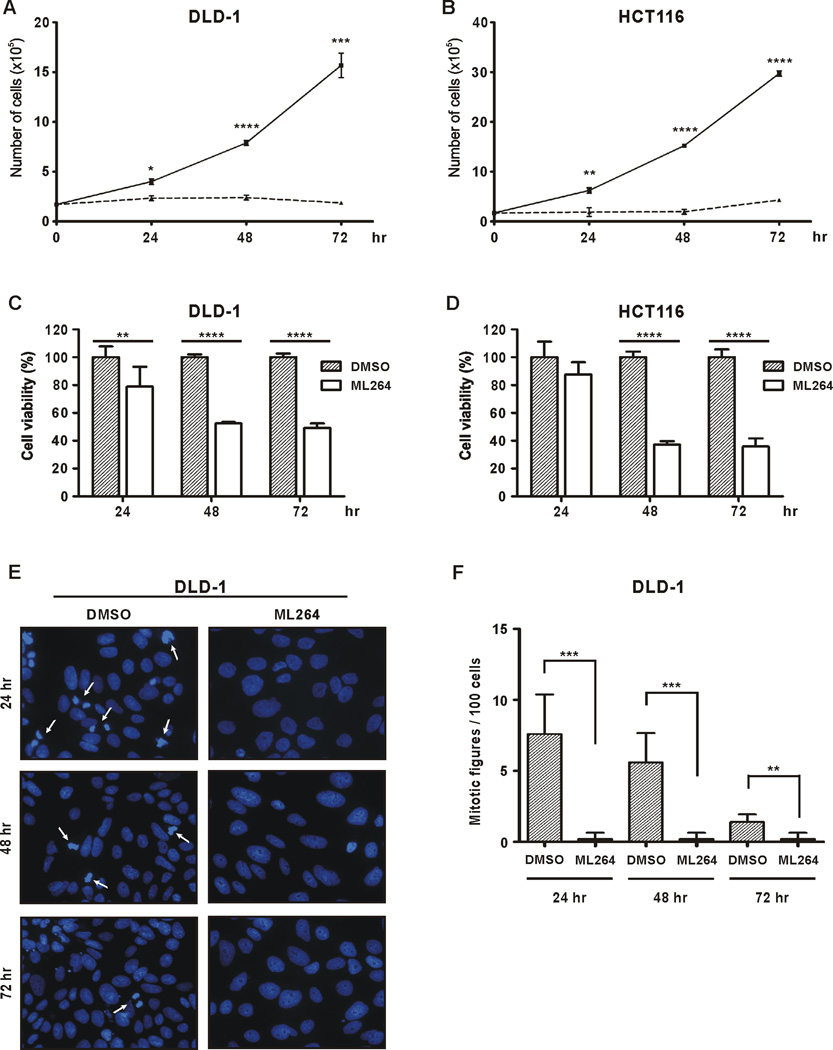
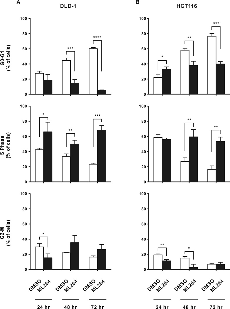
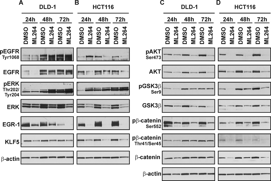
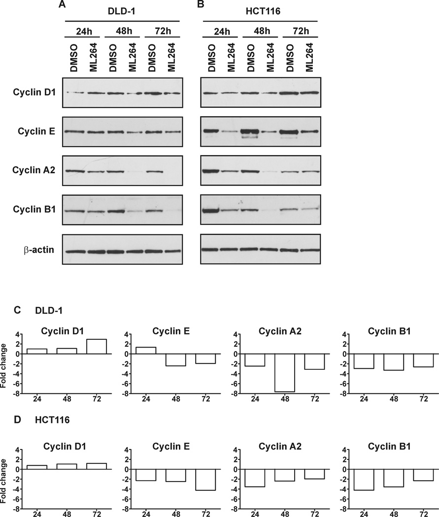
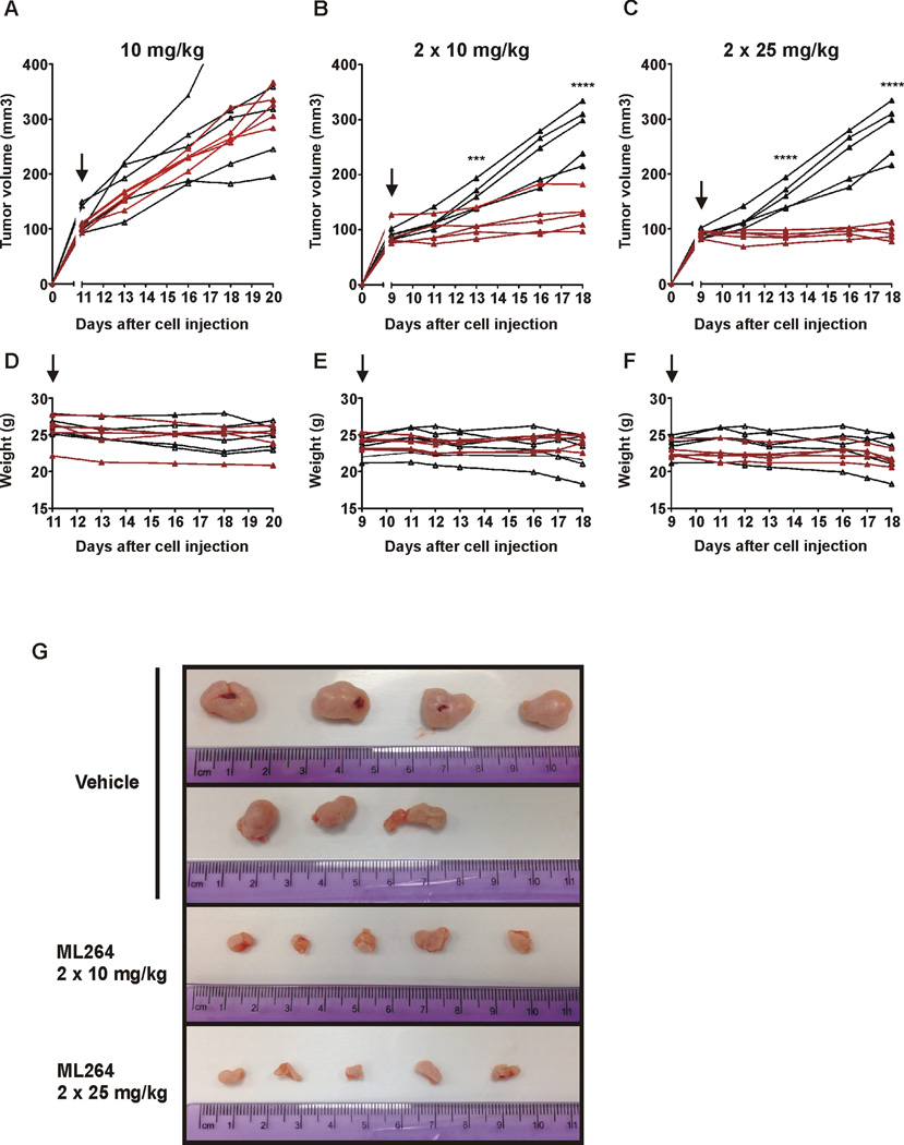
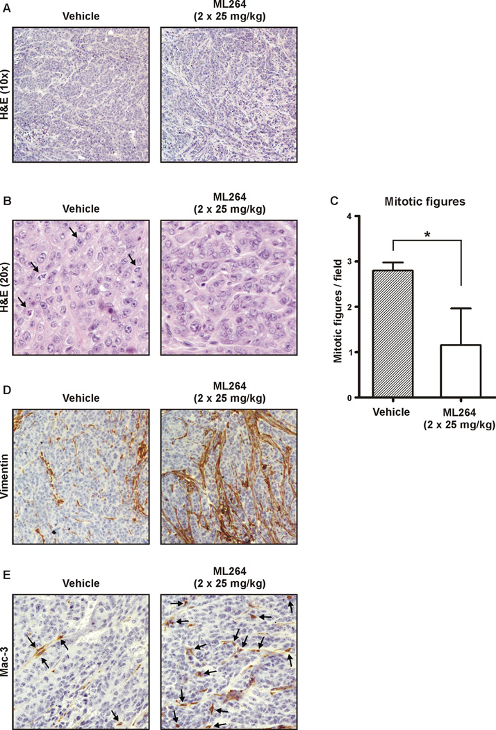
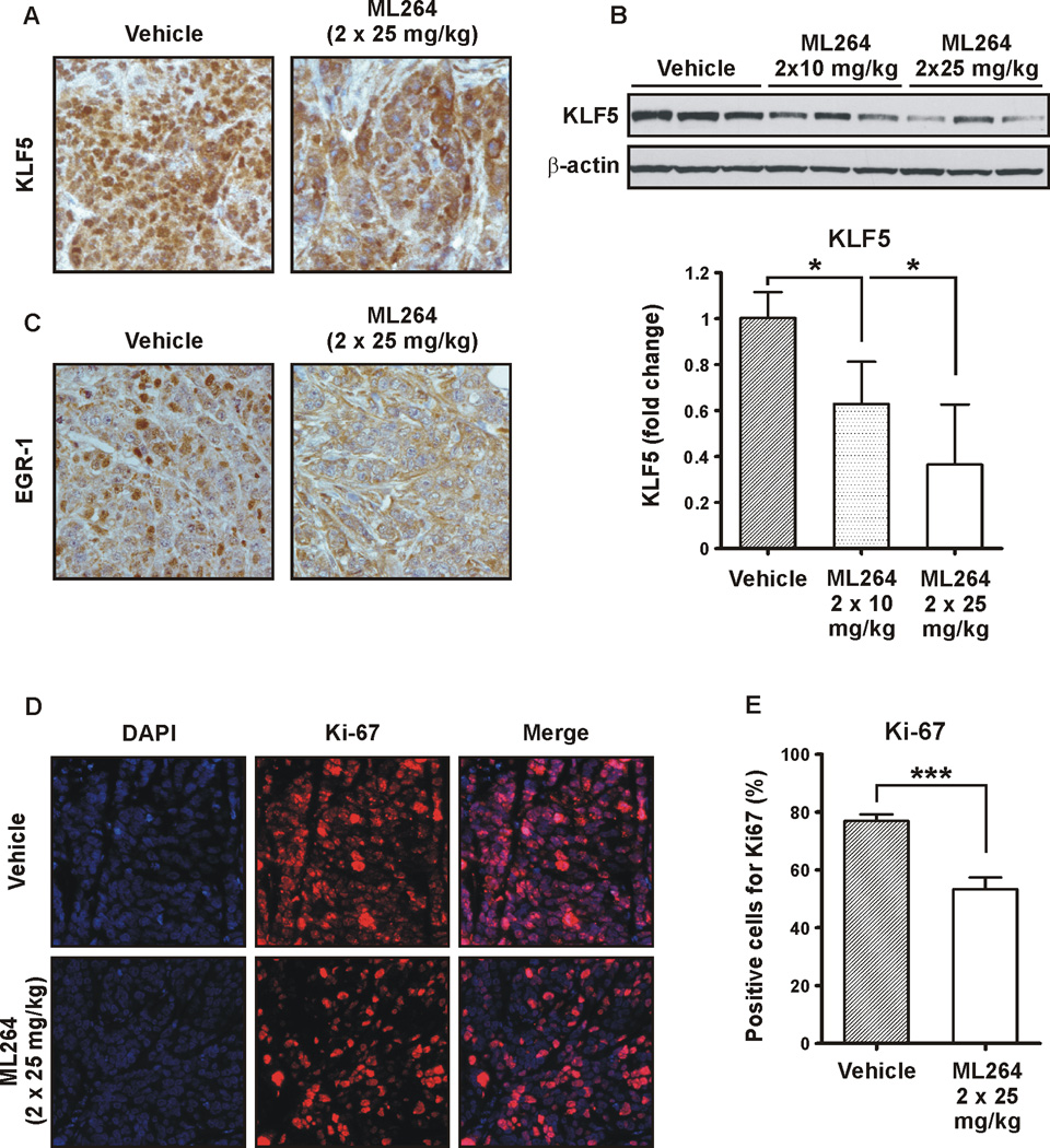
Colorectal cancer is one of the leading causes of cancer mortality in Western civilization. Studies have shown that colorectal cancer arises as a consequence of the modification of genes that regulate important cellular functions. Deregulation of the WNT and RAS/MAPK/PI3K signaling pathways has been shown to be important in the early stages of colorectal cancer development and progression. Krüppel-like factor 5 (KLF5) is a transcription factor that is highly expressed in the proliferating intestinal crypt epithelial cells. Previously, we showed that KLF5 is a mediator of RAS/MAPK and WNT signaling pathways under homeostatic conditions and that it promotes their tumorigenic functions during the development and progression of intestinal adenomas. Recently, using an ultrahigh-throughput screening approach we identified a number of novel small molecules that have the potential to provide therapeutic benefits for colorectal cancer by targeting KLF5 expression. In the current study, we show that an improved analogue of one of these screening hits, ML264, potently inhibits proliferation of colorectal cancer cells in vitro through modifications of the cell-cycle profile. Moreover, in an established xenograft mouse model of colon cancer, we demonstrate that ML264 efficiently inhibits growth of the tumor within 5 days of treatment. We show that this effect is caused by a significant reduction in proliferation and that ML264 potently inhibits the expression of KLF5 and EGR1, a transcriptional activator of KLF5. These findings demonstrate that ML264, or an analogue, may hold a promise as a novel therapeutic agent to curb the development and progression of colorectal cancer.


Conflict of interest statement

There are no conflicts of interest.

Figures

Figure 1
Figure 1. ML264 inhibits proliferation of colorectal cancer cell lines
(A) DLD-1 and (B) HCT116 cells were seeded in 6 well plate format with medium containing DMSO or 10µM ML264. Twenty-four, forty-eight and seventy two hours post-treatment cells were counted using a cell counter. The solid lines represent control and the dotted lines treatment with ML264. Data represent mean ± S.D. (n=3). *p < 0.05, **p < 0.01, ***p < 0.001, ****p < 0.0001. (C) DLD-1 and (D) HCT116 cells were seeded in 96 well plate format with medium containing DMSO or 10µM ML264. Twenty-four, forty-eight and seventy two hours post-treatment cells were analyzed using MTS assay. The measurement of the control (cells with medium and DMSO) was defined as 100% and the results from other measurements were calculated accordingly. Data represent mean ± S.D. (n=6). **p < 0.01, ****p < 0.0001. (E) DLD-1 cells were seeded onto slides with medium containing DMSO or 10µM ML264, fixed and stained with Hoechst (DNA labeling) after 24, 48 and 72 hours of treatment. White arrows marked cells undergoing mitosis. (F) Quantitative representation of (C). Five fields, each with 100 cells were counted. Data represent mean ± S.D. (n=5). **p < 0.01, ***p < 0.001.
Figure 2
Figure 2. ML264 modifies cell cycle progression in colorectal cancer cell lines
(A) DLD-1 and (B) HCT116 cells were seeded in 60 mm plate format with medium containing DMSO or 10µM ML264. Twenty-four, forty-eight and seventy two hours post-treatment cells were collected for cell cycle analysis with propidium iodide. Each experiment was performed in triplicate and data is shown as mean ± S.D. (n=3). *p < 0.05, **p < 0.01, ***p < 0.001, ****p < 0.0001.
Figure 3
Figure 3. Inhibitory effects of ML264 on protein levels of selected components of the MAPK, WNT and PI3K signaling pathways
DLD-1 and HCT116 cells were seeded with medium containing DMSO or 10µM ML264. Twenty-four, forty-eight and seventy two hours post-treatment cells were collected for protein analysis. (A) DLD-1 and (B) HCT116 – Representative Western blots of selected components of MAPK signaling pathway, (C) DLD-1 and (D) HCT116 – Representative Western blots of selected components of PI3K and WNT signaling pathways.
Figure 4
Figure 4. ML264 modulates expression levels of cyclins
DLD-1 and HCT116 cells were seeded with medium containing DMSO or 10µM ML264. Twenty-four, forty-eight and seventy two hours post-treatment cells were collected for protein and RNA analysis. (A) DLD-1 and (B) HCT116 - Representative image of Western blots. (C) DLD-1 and (D) HCT116 – RNA analysis. Fold change is calculated in comparison to vehicle (DMSO) treated cells as described in Materials and Methods section.
Figure 5
Figure 5. ML264 inhibits the growth of DLD-1-derived tumor xenografts in nude mice model
DLD-1 cells were subcutaneously injected into nude mice for the development of xenograft tumors. Mice were treated with vehicle only or with ML264 as follows: daily with 10mg/kg (A, D), twice per day with 10mg/kg (B, E) or twice per day with 25mg/kg (C, F) as detailed in Materials and Methods. Black lines label vehicle-treated mice, and red lines label ML264-treated mice. Black arrows point to the start of injections of vehicle or ML264. Tumor volume is depicted in A–C and mice weight in D–F. The asterisks (***) and (****) indicate a significant difference (p < 0.001) and (p < 0.0001), respectively, between the ML264-treated and vehicle-treated groups. (G) Photographic images of the tumor collected at the end of 10 days treatment as shown in (B) and (C).
Figure 6
Figure 6. Histology and immunohistochemistry of DLD-1-derived tumor xenografts treated with ML264
DLD-1 cells were subcutaneously injected into nude mice for development of xenograft tumors. Mice were treated with vehicle only or with ML264 at 25mg/kg as detailed in Materials and Methods. (A) and (B) Hematoxylin and eosin (H&E)-stained histological images of DLD-1-derived xenografts treated with vehicle (left panel) and treated with ML264 twice per day at 25mg/kg (right panel) for ten days at magnification 10× and 20×, respectively. Black arrows label mitotic figures in panel B. (C) Quantitative representation of (B). Three fields were counted for each condition. Data represent mean ± S.D. (n=3). *p < 0.05. (D) Representative images of immunohistochemistry for vimentin in DLD-1-derived xenografts treated with vehicle (left panel) and treated with ML264 twice per day at 25mg/kg (right panel) for ten days. (E) Representative images of immunohistochemistry for Mac-3 in DLD-1-derived xenografts treated with vehicle (left panel) and treated with ML264 twice per day at 25mg/kg (right panel) for ten days. Black arrows label Mac-3 positive staining.
Figure 7
Figure 7. ML264 treatment reduced the expression levels of KLF5 and EGR1 in DLD-1-derived tumor xenografts
(A) Representative immunohistochemistry staining of KLF5 in DLD-1-derived xenografts treated with vehicle (left panel) and treated with ML264 twice per day at 25mg/kg (right panel) for ten days. (B) Western blot analysis (top panel) and the quantitative analysis (bottom panel) of KLF5 levels in DLD-1-derived xenografts treated with vehicle and treated with ML264 twice per day at 10mg/kg and 25mg/kg for ten days. Results shown from three independent experiments. Data represent mean ± S.D. (n=3). *p < 0.05. (C) Representative immunohistochemistry staining of EGR1 in DLD-1-derived xenografts treated with vehicle (left panel) and treated with ML264 twice per day at 25mg/kg (right panel) for ten days. (D) Immunofluorescence staining of Ki-67 (proliferative marker). Top panel – vehicle treated mice, Bottom panel – ML264-treated mice. (E) Quantitative representation of (D). Three fields were counted for each condition. Data represent mean ± S.D. (n=3). ***p < 0.001.

References

    1. Siegel RL, Miller KD, Jemal A. Cancer statistics, 2015. CA Cancer J Clin. 2015;65:5–29. - PubMed
    1. Arnold CN, Goel A, Blum HE, Boland CR. Molecular pathogenesis of colorectal cancer: implications for molecular diagnosis. Cancer. 2005;104:2035–2047. - PubMed
    1. Mariani F, Sena P, Roncucci L. Inflammatory pathways in the early steps of colorectal cancer development. World journal of gastroenterology : WJG. 2014;20:9716–9731. - PMC - PubMed
    1. Fearon ER. Molecular genetics of colorectal cancer. Annual review of pathology. 2011;6:479–507. - PubMed
    1. Drost J, van Jaarsveld RH, Ponsioen B, Zimberlin C, van Boxtel R, Buijs A, et al. Sequential cancer mutations in cultured human intestinal stem cells. Nature. 2015;521:43–47. - PubMed

Publication types

MeSH terms