. 2016 Feb 15;473(4):423-34.
doi: 10.1042/BJ20150274. Epub 2015 Nov 30.

Autism-associated R451C mutation in neuroligin3 leads to activation of the unfolded protein response in a PC12 Tet-On inducible system

Affiliations

Autism-associated R451C mutation in neuroligin3 leads to activation of the unfolded protein response in a PC12 Tet-On inducible system

Lisa Ulbrich et al. Biochem J. .

Abstract

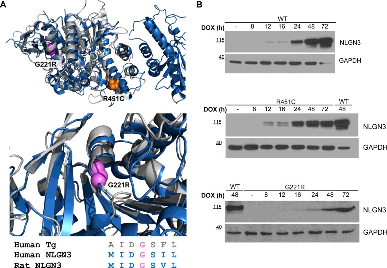
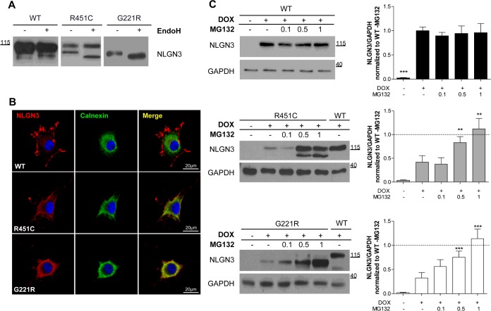
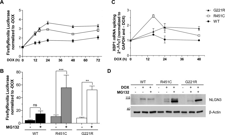
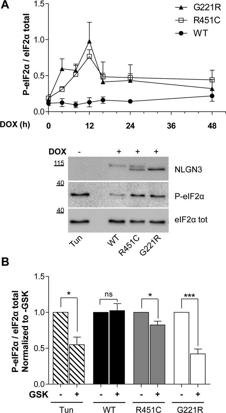
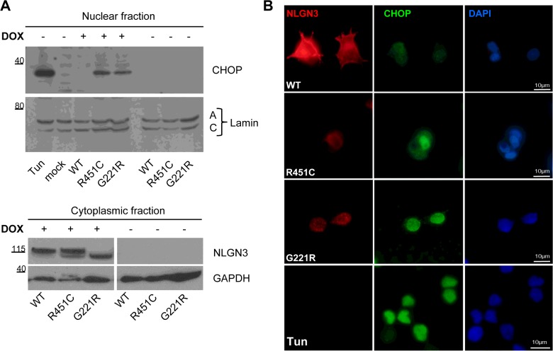
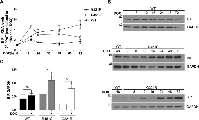
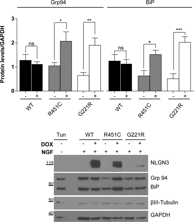
Several forms of monogenic heritable autism spectrum disorders are associated with mutations in the neuroligin genes. The autism-linked substitution R451C in neuroligin3 induces local misfolding of its extracellular domain, causing partial retention in the ER (endoplasmic reticulum) of expressing cells. We have generated a PC12 Tet-On cell model system with inducible expression of wild-type or R451C neuroligin3 to investigate whether there is activation of the UPR (unfolded protein response) as a result of misfolded protein retention. As a positive control for protein misfolding, we also expressed the mutant G221R neuroligin3, which is known to be completely retained within the ER. Our data show that overexpression of either R451C or G221R mutant proteins leads to the activation of all three signalling branches of the UPR downstream of the stress sensors ATF6 (activating transcription factor 6), IRE1 (inositol-requiring enzyme 1) and PERK [PKR (dsRNA-dependent protein kinase)-like endoplasmic reticulum kinase]. Each branch displayed different activation profiles that partially correlated with the degree of misfolding caused by each mutation. We also show that up-regulation of BiP (immunoglobulin heavy-chain-binding protein) and CHOP [C/EBP (CCAAT/enhancer-binding protein)-homologous protein] was induced by both mutant proteins but not by wild-type neuroligin3, both in proliferative cells and cells differentiated to a neuron-like phenotype. Collectively, our data show that mutant R451C neuroligin3 activates the UPR in a novel cell model system, suggesting that this cellular response may have a role in monogenic forms of autism characterized by misfolding mutations.

Keywords: ER stress; autism; molecular chaperones; neuroligin; protein misfolding; unfolded protein response.


Figures

Figure 1
Figure 1. Structure and time course of expression of NLGN3 in inducible PC12 Tet-On clones
(A) Upper panel: overlay of the three dimensional structure of the α/β-hydrolase domain of NLGN4 (blue, PDB code 3BE8) and the homology model of the cholinesterase-like domain of Tg (grey), with the location of the R451C (orange) and G221R (magenta) mutations. Overlay was obtained using the program PyMOL (http://www.pymol.org). The Swiss Model Server (http://swissmodel.expasy.org/) was used to obtain structural template for the cholinesterase-like domain of Tg (residue 2192 to residue 2719). Lower panel: magnification of the homologous region between NLGN4 and Tg where G221R/G2300R are located. Sequence alignment shows that the glycine residue is highly conserved in NLGN3 and Tg. (B) NLGN3 expression in PC12 cells after DOX treatment (0–72 h). WT lysates at 48 h from DOX were used as a reference for migration and band intensity across the three panels. Molecular masses are indicated in kDa.
Figure 2
Figure 2. Localization and degradation of NLGN3 in inducible PC12 Tet-On clones
(A) EndoH treatment of lysates from cells expressing WT, R451C and G221R NLGN3. The image presents different parts of the same Western blot. (B) NLGN3 localization in PC12 cells differentiated for 5 days with NGF. WT NLGN3 (FLAG, red) did not show any co-localization with calnexin (green), in contrast with the R451C and G221R mutant proteins showing partial and complete co-localization (yellow). DAPI (blue) was used to stain the nuclei. (C) Left panels: Western blot analysis of NLGN3 protein levels after 48 h of DOX treatment and 24 h of MG132 (0.1, 0.5 and 1μM). GAPDH was used as loading control. Histograms on the right show NLGN3/GAPDH densitometric analysis normalized to WT NLGN3 levels in the absence of MG132. Statistical analysis comparing treated and untreated conditions within each clone (n=4; **P<0.01, ***P<0.001). Molecular masses are indicated on the blots in kDa.
Figure 3
Figure 3. Activation of the UPR transcriptional factors
(A) PC12 cells were transfected with the luciferase reporter vectors after inducing NLGN3 synthesis with DOX (from 0 to 72 h). For each clone, the ratio of firefly/Renilla luciferase was normalized to the −DOX condition at each of the time points. Results are means±S.E.M. for six independent experiments. Mutants compared with WT values, for all time points: ***P<0.001, except for R451C at 48 h, *P<0.05 and 72 h, **P<0.01. R451C compared with G221R at 24 h and 48 h: **P<0.01. (B) NLGN3 synthesis was induced for 48 h with DOX and 0.5 μM MG132 was added for the last 24 h. Results are means±S.E.M. for five independent experiments (***P<0.001 and **P<0.01). (C) NLGN3 synthesis was induced, from 0 to 48 h, and the splicing of XBP1 was quantified by real-time RT–PCR. Changes in XBP1s were calculated relative to the 18S housekeeping gene and normalized to the −DOX condition. For each time point, statistical relevance of four independent experiments was calculated comparing NLGN3 mutants with WT. A peak value of XBP1s was observed at 16 h from DOX induction in the R451C NLGN3 cells (R451C compared with WT, ***P<0.001; R451C compared with G221R, **P<0.01), whereas G221R NLGN3 overexpression caused a slower but steady XBP1 splicing increase (*P<0.05 at 24 h and **P<0.01 at 48 h). XBP1 splicing in WT NLGN3 cells was unchanged over time. (D) NLGN3 protein levels were quantified after DOX induction of NLGN3 synthesis for 16 h in the presence or absence of MG132 (0.5 μM). The Western blot is representative of four independent experiments. Molecular masses are indicated in kDa.
Figure 4
Figure 4. PERK branch activation
(A) Upper panel: time course densitometric analysis of Western blot for p-eIF2α, from 0 to 48 h from DOX treatment, normalized to total eIF2α levels. Significant differences were observed for R451C and G221R NLGN3 mutant clones at 12 h from DOX treatment (**P<0.01 and ***P<0.001 respectively) in comparison with WT. Mean±S.E.M. values were from four independent experiments. Lower panel: representative Western blot for p-eIF2α and total eIF2α from PC12 NLGN3 Tet-On cells at 12 h after DOX administration. Molecular masses are indicated in kDa. (B) Densitometry of p-eIF2α normalized to total eIF2α from four independent experiments on PC12 cells at 12 h from DOX induction, without or with 50 nM GSK2606414 treatment. Mean±S.E.M. values were from four independent experiments (*P<0.05, **P<0.01, ***P<0.001).
Figure 5
Figure 5. Expression levels of the UPR target CHOP
(A) Nuclear and cytoplasmic fractions extracted from PC12 NLGN3 cells at 24 h after DOX administration. Upper panel: Western blot detection of CHOP. Parental PC12 cells (mock) and the −DOX conditions were used as negative controls and tunicamycin was used as a positive control. Nuclear fraction was normalized to lamin A/C protein. The Western blot is representative of four independent experiments. Lower panel: NLGN3 detection in the cytoplasmic protein fraction. GAPDH was used as a loading control. Image presents two different parts of the same Western blot. Molecular masses are indicated in kDa. (B) Differentiated PC12 NLGN3 cells stained with the anti-CHOP antibody (green) and anti-FLAG for detection of NLGN3 (red). Tunicamycin-treated parental PC12 cells were used as a positive control (lower panel).
Figure 6
Figure 6. Up-regulation of the UPR target BiP
(A) NLGN3 was induced from 0 to 72 h by DOX and BiP mRNA levels were quantified. Time point values were normalized to −DOX, for each clone, and statistically compared with WT. Results are means±S.E.M. for four independent experiments. At 16 h from DOX, a robust increase in BiP mRNA is observed in the NLGN3 R451C (**P<0.01) with respect to WT and is still significant at 48 h (*P<0.05). The G221R mutant protein caused an increase over the time becoming gradually significant from 24 to 72 h (*P<0.05 at 24 h, **P<0.01 at 48 h, ***P<0.001 at 72 h). BiP mRNA levels showed a flat trend for the WT NLGN3 clone. Comparison between R451C and G221R revealed significant differences between the two mutants at 72 h, *P<0.05. (B) Representative time course of BiP protein levels after DOX treatment from 0 to 72 h. Molecular masses are indicated in kDa. (C) Densitometry of BiP normalized to GAPDH levels was calculated after inducing NLGN3 synthesis for 24 h (n=4). Statistical analysis compared the +DOX with the −DOX conditions (R451C *P<0.05, G221R **P<0.01).
Figure 7
Figure 7. Levels of Grp94 and BiP in differentiated PC12 NLGN3 Tet-On clones
Upper panel: densitometry of Grp94 (left) and BiP (right) levels in mutant and WT cells treated with NGF and DOX for 72 h (n=3). Statistical analysis compared values of the +DOX condition with the −DOX condition (*P<0.05, **P<0.01, ***P<0.001). Lower panel: representative Western blot of NGF-differentiated PC12 cells for the detection of Grp94 and BiP using the anti-KDEL antibody. βIII-Tubulin was used as a marker for differentiation and GAPDH was used as a loading control. Undifferentiated parental PC12 cells treated with tunicamycin were used as positive control. Molecular masses are indicated in kDa.

Similar articles

Cited by

References

    1. Pinto D., Delaby E., Merico D., Barbosa M., Merikangas A., Klei L., Thiruvahindrapuram B., Xu X., Ziman R., Wang Z., et al. Convergence of genes and cellular pathways dysregulated in autism spectrum disorders. Am. J. Hum. Genet. 2014;94:677–694. doi: 10.1016/j.ajhg.2014.03.018. - DOI - PMC - PubMed
    1. Südhof T.C. Neuroligins and neurexins link synaptic function to cognitive disease. Nature. 2008;455:903–911. doi: 10.1038/nature07456. - DOI - PMC - PubMed
    1. Jamain S., Quach H., Betancur C., Rastam M., Colineaux C., Gillberg I.C., Soderstrom H., Giros B., Leboyer M., Gillberg C., Bourgeron T. Mutations of the X-linked genes encoding neuroligins NLGN3 and NLGN4 are associated with autism. Nat. Genet. 2003;34:27–29. doi: 10.1038/ng1136. - DOI - PMC - PubMed
    1. Comoletti D., De Jaco A., Jennings L.L., Flynn R.E., Gaietta G., Tsigelny I., Ellisman M.H., Taylor P. The Arg451Cys-neuroligin-3 mutation associated with autism reveals a defect in protein processing. J. Neurosci. 2004;24:4889–4893. doi: 10.1523/JNEUROSCI.0468-04.2004. - DOI - PMC - PubMed
    1. Chih B., Afridi S.K., Clark L., Scheiffele P. Disorder-associated mutations lead to functional inactivation of neuroligins. Hum. Mol. Genet. 2004;13:1471–1477. doi: 10.1093/hmg/ddh158. - DOI - PubMed

Publication types

MeSH terms