Case Reports
. 2015 Nov 30:2015:bcr2015212970.
doi: 10.1136/bcr-2015-212970.

Gliosarcoma with neuroaxis metastases

Affiliations
Case Reports

Gliosarcoma with neuroaxis metastases

Rui Ramos et al. BMJ Case Rep. .

Abstract

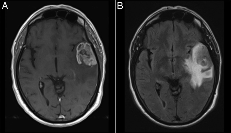
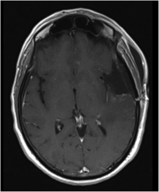
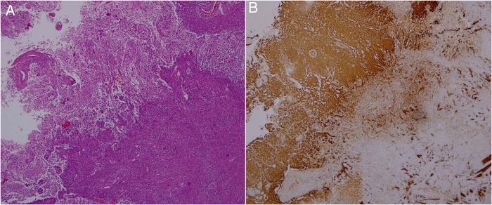
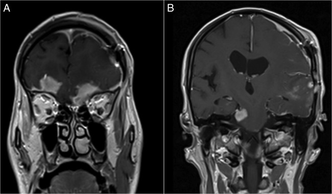
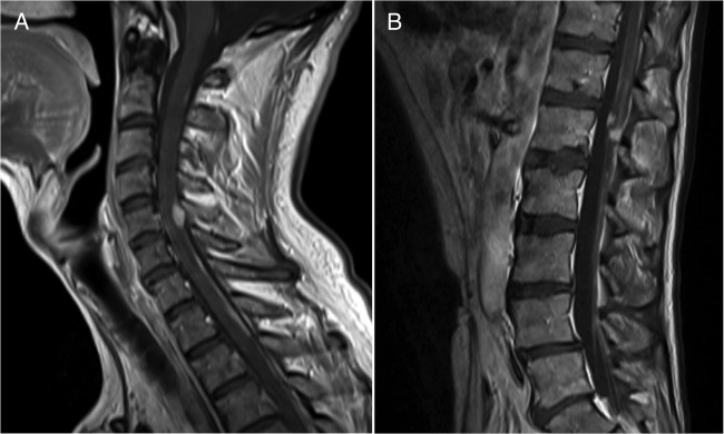
Gliosarcomas are rare tumours of the central nervous system, with a well-known capacity for metastasis. When they metastasise, the dissemination occurs more frequently via the haematogenous route to extraneural sites. Metastasis-spread through the cerebrospinal fluid is extremely rare. We present the case of a 58-year-old man who underwent a gross total resection of a lesion in the left temporal lobe. The histological findings revealed a gliosarcoma and the patient received radiotherapy followed by chemotherapy. Seven months after surgery, while the patient remained neurologically intact, brain and spinal cord MRI revealed tumour recurrence and neuroaxis metastases through the traffic routes of the cerebrospinal fluid. The patient died 8 months after the diagnosis. A PubMed search regarding metastatic gliosarcoma up to June 2015 was also carried out. To the best of our knowledge, this is the first case report of gliosarcoma metastases to the brain and spinal cord leptomeninges.


Figures

Figure 1
Figure 1
Preoperative brain MRIs. (A) Gadolinium-enhanced T1-weighted axial image showing a lesion in the left temporal lobe with heterogeneous contrast uptake. (B) Fluid-attenuated inversion recovery axial image showing tumoural infiltration/oedema in the surrounding brain parenchyma.
Figure 2
Figure 2
Postoperative brain MRI. Gadolinium-enhanced T1-weighted axial image demonstrating radical tumour excision.
Figure 3
Figure 3
Histological tissue sections. (A) H&E showing a biphasic tumour with glial (left) and fusiform (right) cells (×40 original magnification). (B) Glial fibrillar acidic protein with strong immunoreactivity in the glial component (left) and virtually no staining in the mesenchymal tissue (right; ×40 original magnification).
Figure 4
Figure 4
MRIs of the control brain at 7 months after craniotomy. Gadolinium-enhanced T1-weighted coronal images showing: (A) leptomeningeal spread with multiple parenchyma and meningeal deposits, (B) one of which involves the right trigeminal nerve.
Figure 5
Figure 5
Vertebrospinal MRI at 7 months after craniotomy. Gadolinium-enhanced T1-weighted sagittal images revealing: (A) retroclival enhancement, diffuse meningeal spread, and nodular lesions in the C5 and (B) along the cauda equina nerve roots, indicative of ‘drop’ metastases.

References

    1. Demirci S, Akalin T, Islekel S et al. . Multiple spinal metastases of cranial gliosarcoma: a case report and review of the literature. J Neurooncol 2008;88:199–204. 10.1007/s11060-008-9550-4 - DOI - PubMed
    1. Han SJ, Yang I, Ahn BJ et al. . Clinical characteristics and outcomes for a modern series of primary gliosarcoma patients. Cancer 2010;116:1358–66. 10.1002/cncr.24857 - DOI - PubMed
    1. Guney Y, Hiçsönmez A, Yilmaz S et al. . Gliosarcoma: a study of four cases. Rare Tumors 2010;2:e37 10.4081/rt.2010.e37 - DOI - PMC - PubMed
    1. Biswas A, Kumar N, Kumar P et al. . Primary gliosarcoma-clinical experience from a regional cancer centre in north India. Br J Neurosurg 2011;25:723–9. 10.3109/02688697.2011.570881 - DOI - PubMed
    1. Han SJ, Yang I, Tihan T et al. . Primary gliosarcoma: key clinical and pathologic distinctions from glioblastoma with implications as a unique oncologic entity. J Neurooncol 2010;96:313–20. 10.1007/s11060-009-9973-6 - DOI - PMC - PubMed

Publication types

MeSH terms