

Dellen and corneal perforation after bilateral pterygium excision in a patient with no risk factors
- PMID: 26621907
- PMCID: PMC4680279
- DOI: 10.1136/bcr-2015-213319
Dellen and corneal perforation after bilateral pterygium excision in a patient with no risk factors
Abstract
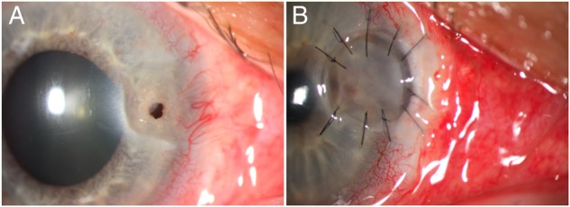
Corneal perforation associated with dellen is a rare but serious complication of a primary pterygium excision. In this case report, we describe a 60-year-old man who underwent a corneal perforation in the centre of corneal dellen in his right eye and corneal dellen in his left eye after the surgical treatment of a bilateral pterygium with a bare sclera technique without adjunctive therapy. He was successfully treated with lamellar keratoplasty in his right eye and a conservative approach in his left eye, consisting of the use of artificial tears, antibiotic ointment and a patch. The clinical evidence from this brief interventional case report indicates that topical lubricants are proper therapy for corneal dellen. However, if corneal perforation is observed, lamellar keratoplasty is a good option.
2015 BMJ Publishing Group Ltd.
Figures
References
-
- Fuchs E. Ueber Dellen in der Cornea. Graefe's Arch Ophthalmol 1911;78:82–92. 10.1007/BF02124899 - DOI
Publication types
MeSH terms
LinkOut - more resources
Full Text Sources
Other Literature Sources
Medical