Review
. 2015 Dec;24(138):642-52.
doi: 10.1183/16000617.0062-2015.

Right heart catheterisation: best practice and pitfalls in pulmonary hypertension

Affiliations
Review

Right heart catheterisation: best practice and pitfalls in pulmonary hypertension

Stephan Rosenkranz et al. Eur Respir Rev. 2015 Dec.

Abstract

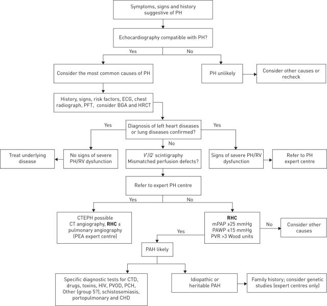
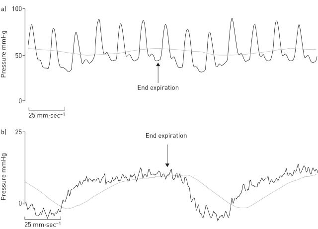
Right heart catheterisation (RHC) plays a central role in identifying pulmonary hypertension (PH) disorders, and is required to definitively diagnose pulmonary arterial hypertension (PAH). Despite widespread acceptance, there is a lack of guidance regarding the best practice for performing RHC in clinical practice. In order to ensure the correct evaluation of haemodynamic parameters directly measured or calculated from RHC, attention should be drawn to standardising procedures such as the position of the pressure transducer and catheter balloon inflation volume. Measurement of pulmonary arterial wedge pressure, in particular, is vulnerable to over- or under-wedging, which can give rise to false readings. In turn, errors in RHC measurement and data interpretation can complicate the differentiation of PAH from other PH disorders and lead to misdiagnosis. In addition to diagnosis, the role of RHC in conjunction with noninvasive tests is widening rapidly to encompass monitoring of treatment response and establishing prognosis of patients diagnosed with PAH. However, further standardisation of RHC is warranted to ensure optimal use in routine clinical practice.


Conflict of interest statement

Conflict of interest: Disclosures can be found alongside the online version of this article at err.ersjournals.com

Figures

FIGURE 1
FIGURE 1
Diagnostic algorithm for pulmonary arterial hypertension (PAH). PH: pulmonary hypertension; PFT: pulmonary function testing; BGA: blood gas analysis; HRCT: high-resolution computed tomography; RV: right ventricular; V′/Q′: ventilation/perfusion; CTEPH: chronic thromboembolic pulmonary hypertension; CT: computed tomography; RHC: right heart catheterisation; PEA: pulmonary endarterectomy; mPAP: mean pulmonary arterial pressure; PAWP: pulmonary arterial wedge pressure; PVR: pulmonary vascular resistance; CTD: connective tissue disease; PVOD: pulmonary veno-occlusive disease; PCH: pulmonary capillary haemangiomatosis; CHD: congenital heart disease. Reproduced and modified from [4] with permission from the publisher.
FIGURE 2
FIGURE 2
Best practice recommendations for right heart catheterisation: pressure transducer and zeroing [3, 20]. The joint task force of the European Society of Cardiology and the European Respiratory Society recommends setting the pressure transducer to zero at the mid-thoracic line (with a suggested reference point defined by the intersection of the frontal plane at the mid-thoracic level, the transverse plane at the level of fourth anterior intercostal space, and the midsagittal plane [20]) in a supine patient halfway between the anterior sternum and the bed surface [4]. Reproduced from [20] with permission from the publisher.
FIGURE 3
FIGURE 3
Representative pressure tracings of a) pulmonary arterial pressure and b) pulmonary arterial wedge pressure (PAWP). Both recordings display respiratory variations, and the correct measurements should be made at the end of the expiratory phase (arrows). For measurement of PAWP (b), the digital read is usually obtained automatically and averages over the inspiration and expiration, with a resultant value of 4 mmHg. The correct value is indicated by the arrow marking the end-expiratory phase when the respiratory system is at functional residual capacity and is 10 mmHg.
FIGURE 4
FIGURE 4
Potential for misclassification of pulmonary hypertension with reliance on end-expiratory pulmonary arterial wedge pressure (PAWP). Distribution of PAWP at end exhalation versus as calculated by the respiratory mean according to clinical phenotype. Larger circles represent identical observations in multiple patients. Blue circles above 15 mmHg represent phenotypically pre-capillary patients who would have been misclassified as having pulmonary venous hypertension using end-expiratory PAWP. Reproduced from [30] with permission from the publisher.

Similar articles

Cited by

References

    1. Chatterjee K. The Swan-Ganz catheters: past, present, and future. A viewpoint. Circulation 2009; 119: 147–152. - PubMed
    1. Nossaman BD, Scruggs BA, Nossaman VE, et al. . History of right heart catheterization: 100 years of experimentation and methodology development. Cardiol Rev 2010; 18: 94–101. - PMC - PubMed
    1. Hoeper MM, Bogaard HJ, Condliffe R, et al. . Definitions and diagnosis of pulmonary hypertension. J Am Coll Cardiol 2013; 62: Suppl., D42–D50. - PubMed
    1. Galiè N, Humbert M, Vachiery JL, et al. . 2015 ESC/ERS Guidelines for the diagnosis and treatment of pulmonary hypertension. Eur Respir J 2015; 46: 903–975. - PubMed
    1. Simonneau G, Gatzoulis MA, Adatia I, et al. . Updated clinical classification of pulmonary hypertension. J Am Coll Cardiol 2013; 62: Suppl., D34–D41. - PubMed

Publication types

MeSH terms