. 2015 Sep;10(3):1202-1206.
doi: 10.3892/etm.2015.2614. Epub 2015 Jul 3.

Treatment of nasal microcystic adnexal carcinoma with an expanded rotational forehead skin flap: A case report and review of the literature

Affiliations

Treatment of nasal microcystic adnexal carcinoma with an expanded rotational forehead skin flap: A case report and review of the literature

Rong-Rong Zhou et al. Exp Ther Med. 2015 Sep.

Abstract

Microcystic adnexal carcinoma (MAC) is a rare and locally aggressive adenocarcinoma with low-grade malignancy. The present study describes the first reported case and treatment of a Chinese male with a MAC located on the nasal dorsum and nosewing. A 44-year-old man presented with a nasal deformity caused by local repeated infections following an accidental injury to the nose 20 years previously. The nose had been injured by a brick, and treatment at a local hospital 12 years previously had resulted in a nasal scar and a gradually enlarging mass. A physical examination revealed a hypertrophic deformity of the nose and an indurated scar plaque, measuring 2.0×2.0 cm, on the nasal dorsum and nosewing. Microscopic examination revealed a tumor consisting of solid cell nests and a cystic structure with a capsular space. In addition, ductal cells of an adnexal cell origin were visible in the outer epithelium. The medial portion exhibited a microductal structure and invasion of deeper tissues without evident atypia. The tumor cells presented normal nuclear to cytoplasmic ratios and minimal mitotic activity. Pathological examination verified that the tumor was a MAC of low-grade malignancy. A complete surgical resection was performed via Mohs micrographic surgery (MMS), and reconstruction was achieved using an expanded rotational forehead skin flap. No tumor recurrence was detected after a three-year follow-up period. Therefore, for effective treatment of similar MAC cases, complete surgical resection using MMS is recommended, and successful reconstruction may be achieved using an expanded skin flap.

Keywords: Mohs micrographic surgery; microcystic adnexal carcinoma; rotational forehead skin flap.


Figures
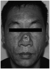
Figure 1.
Figure 1.
Macroscopic appearance of the tumor. The patient presented with a tumor that was similar in appearance to a depressed scar without ulceration or clinical symptoms.
Figure 2.
Figure 2.
Generation of an expanded forehead skin flap. (A) A forehead tissue expander was implanted; (B) extensive resection of the tumor was conducted; (C and D) reconstruction was performed using the expanded rotational forehead skin flap.
Figure 3.
Figure 3.
Reconstruction with the expanded forehead skin flap, as shown from the (A) anterior position, (B) right anterior oblique position and (C) right lateral position.
Figure 4.
Figure 4.
Flap separation. (A) Abscise of the flap at the distal; (B) suture of the flap incision at the nasal root; (C) suture of the flap incision at the forehead.
Figure 5.
Figure 5.
Appearance of the reconstruction following removal of the stitches from an (A) anterior position, (B) right anterior oblique position and (C) right lateral position. Reconstruction of the defect resulted in normal function and esthetics.
Figure 6.
Figure 6.
Histopathological observations. (A) Exterior portion of the tumor exhibited solid cell nests and cystic structures (shown by arrows) with a capsular space (magnification, x40). (B) Ductal cells (shown by arrows) were visible in the exterior portion, appearing as clear cells originating from adnexal cells (magnification, x200). (C) Medial tumor portion comprised a microductal structure (as shown by arrow) formed by ductal epithelial cells, with a tendency to invade the deeper tissues without evident atypia (magnification, x200). (D) Tumor cells in the medial portion exhibited a normal nuclear to cytoplasmic ratio (as shown by arrow), and minimal mitotic activity (magnification, x200). (E) Deeper tumor tissue exhibited marked interstitial sclerosis and collagenization (as shown by arrow; magnification, x100).

References

    1. Goldstein DJ, Barr RJ, Santa Cruz DJ. Microcystic adnexal carcinoma: A distinct clinicopathologic entity. Cancer. 1982;50:566–572. doi: 10.1002/1097-0142(19820801)50:3<566::AID-CNCR2820500330>3.0.CO;2-Q. - DOI - PubMed
    1. Yu JB, Blitzblau RC, Patel SC, Decker RH, Wilson LD. Surveillance, Epidemiology, and End Results (SEER) database analysis of microcystic adnexal carcinoma (sclerosing sweat duct carcinoma) of the skin. Am J Clin Oncol. 2010;33:125–127. - PubMed
    1. Bier-Laning cm, Hom DB, Gapany M, Manivel JC, Duvall AJ., III Microcystic adnexal carcinoma: Management options based on long-term follow-up. Laryngoscope. 1995;105:1197–1201. doi: 10.1288/00005537-199511000-00011. - DOI - PubMed
    1. Lipper S, Peiper SC. Sweat gland carcinoma with syringomatous features: A light microscopic and ultrastructural study. Cancer. 1979;44:157–163. doi: 10.1002/1097-0142(197907)44:1<157::AID-CNCR2820440127>3.0.CO;2-4. - DOI - PubMed
    1. Glatt HJ, Proia AD, Tsoy EA, Fetter BF, Klintworth GK, Neuhaus R, Font RL. Malignant syringoma of the eyelid. Ophthalmology. 1984;91:987–990. doi: 10.1016/S0161-6420(84)34204-X. - DOI - PubMed

LinkOut - more resources