. 2015 Oct;10(4):1327-1333.
doi: 10.3892/etm.2015.2660. Epub 2015 Jul 27.

Rapamycin-induced autophagy activity promotes bone fracture healing in rats

Affiliations

Rapamycin-induced autophagy activity promotes bone fracture healing in rats

G E Yang et al. Exp Ther Med. 2015 Oct.

Erratum in

Abstract

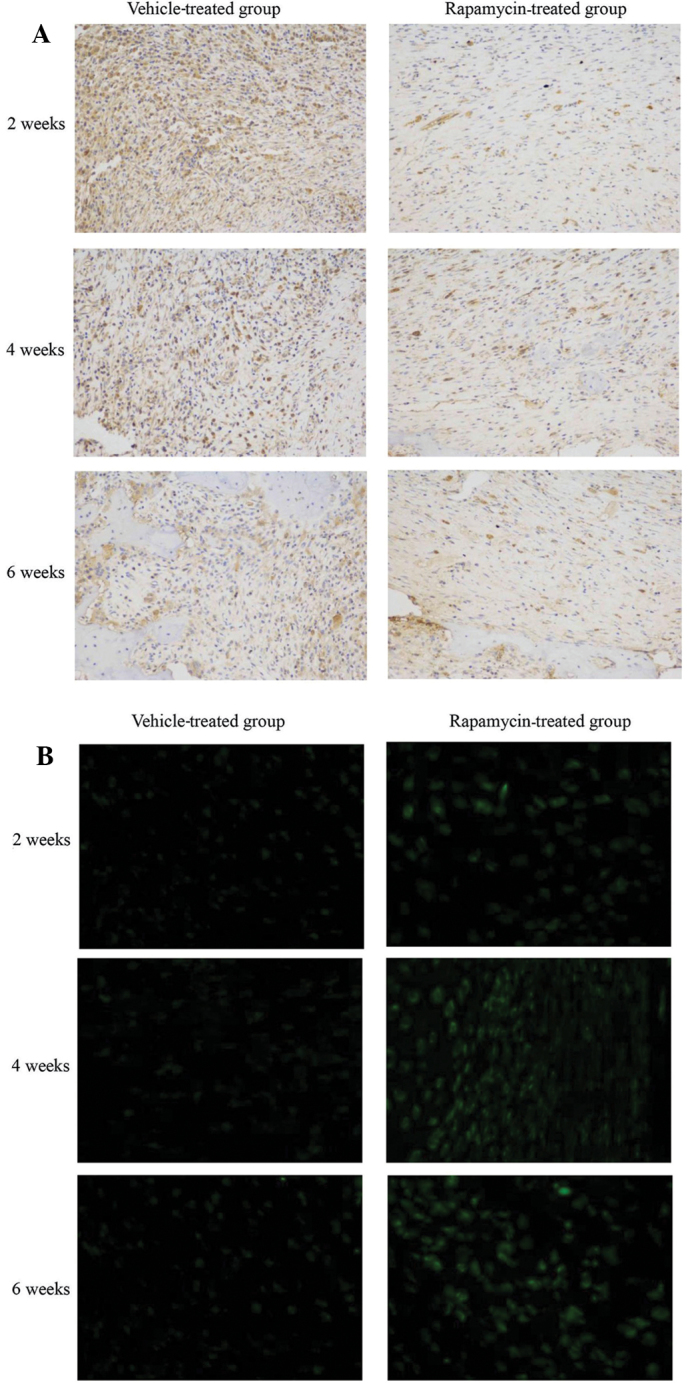
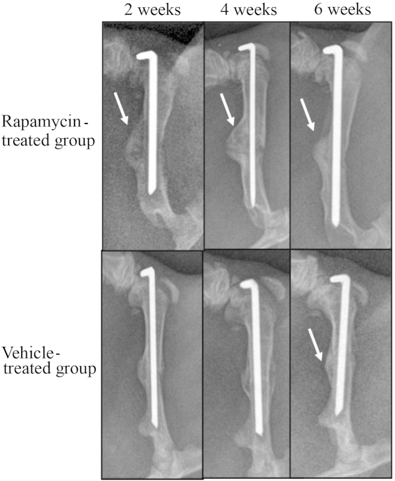
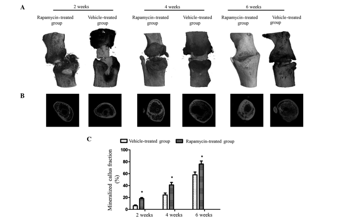
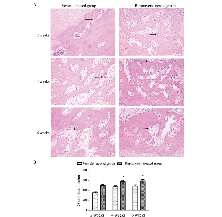
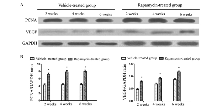
Autophagy is a crucial mediating process for normal bone cell function and metabolism in physiology or pathology. Rapamycin has been demonstrated to induce the autophagy pathway by inhibiting the mammalian target of rapamycin (mTOR) pathway. However, the contribution of autophagy in orthopedic diseases is rarely reported. The aim of the present study was to evaluate the capacity of pharmacologically induced autophagy to modify disease function in a rat model of bone fracture. A femur fracture model was established via surgery in adult male Sprague-Dawley rats. Rapamycin (n=63 rats) or dimethyl sulfoxide (DMSO) vehicle control (n=63 rats) was administered intraperitoneally for 2, 4 and 6 weeks, and 21 randomly selected rats were sacrificed in each group at each time point. X-ray micro-computed tomography and hematoxylin and eosin staining were used to evaluate the extent of fracture healing in each group. The effects of rapamycin on autophagy, mTOR signaling and the expression levels of vascular endothelial growth factor (VEGF) and proliferating cell nuclear antigen (PCNA) were analyzed using immunohistochemistry, immunofluorescence staining and western blot analysis. Rapamycin affected the mTOR signaling pathway in rats following fracture, as indicated by the inhibition of the phosphorylation of ribosomal protein S6, a target of mTOR, and activation of microtubule-associated protein 2 light chain 3, a key marker of autophagy. Histomorphometry and image examination indicated that the number of osteoblasts in each section was significantly (P<0.01) increased in the rapamycin group compared with the control group, and this was associated with a significant (P<0.05) increase in mineralized callus fraction. Furthermore, rapamycin treatment increased the expression levels of VEGF and PCNA in the rat callus tissue. These results suggest that rapamycin may serve a beneficial function in fracture healing, and that the underlying mechanism may involve the activation of autophagy.

Keywords: autophagy; bone fracture healing; rapamycin.


Figures

Figure 1.
Figure 1.
Systemic administration of rapamycin modulates the mammalian target of rapamycin signaling pathway and autophagy in a rat fracture model. Calluses from rats were collected at 2, 4 and 6 weeks post-fracture after treatment with rapamycin or the vehicle (n=12 per group). (A) Sections were analyzed using immunohistochemistry for phosphorylation of ribosomal protein S6. (B) Sections were analyzed by immunofluorescence for light chain 3-II (magnification, ×200).
Figure 2.
Figure 2.
Effect of rapamycin on the size and mineralization of fracture calluses during bone repair. Representative X-ray images of fracture calluses from rapamycin and vehicle-treated rats at 2, 4 and 6 weeks post-fracture. Arrow indicates density of mineralization with fracture calluses.
Figure 3.
Figure 3.
Influence of rapamycin on fracture callus mineralization and remodeling. Bone fracture global imaging was used to rebuild bone structure by micro-computed tomography (micro-CT). All fracture callus sites from rapamycin- and vehicle-treated rats were harvested at 2, 4 and 6 weeks post-fracture (n=15 per group). (A and B) Global image and transverse section of fracture calluses were scanned and graphically re-constructed using micro-CT. (C) Mineralized callus fraction was analyzed using micro-CT. Values are presented as the mean ± standard deviation. *P<0.05 vs. the vehicle-treated group.
Figure 4.
Figure 4.
Rapamycin increase the number of osteoblasts in the rat callus. (A) Calluses section from Sprague-Dawley rats at 2, 4 and 6 weeks post-fracture under treatment with rapamycin or vehicle were stained with hematoxylin and eosin (n=12 per group; magnification, ×200). Arrow indicates an osteoblast in the callus. (B) Quantitative analysis of osteoblast number indicated a significant increase in cellularity after rapamycin treatment compared with the vehicle treatment. Values are presented as the mean ± standard deviation. *P<0.01 vs. vehicle-treated group.
Figure 5.
Figure 5.
Effects of rapamycin on PCNA and VEGF expression in the callus. (A) Western blot analysis of PCNA and VEGF expression levels in the rat callus at 2, 4 and 6 weeks post-fracture. (B) Quantitative comparison of PCNA and VEGF expression levels between vehicle-treated and rapamycin groups, expressed as a ratio of GAPDH, used as loading control (n=12 per group). Values are presented as the mean ± standard deviation. *P<0.05 vs. vehicle-treated group. PCNA, proliferating cell nuclear antigen; VEGF, vascular endothelial growth factor; GAPDH, glyceraldehyde 3-phosphate dehydrogenase.

References

    1. Einhorn T. Enhancement of fracture-healing. J Bone Joint Surg Am. 1995;77:940–956. - PubMed
    1. O'Neill KR, Stutz CM, Mignemi NA, et al. Micro-computed tomography assessment of the progression of fracture healing in mice. Bone. 2012;50:1357–1367. doi: 10.1016/j.bone.2012.03.008. - DOI - PubMed
    1. Mizushima N, Levine B, Cuervo AM, Klionsky DJ. Autophagy fights disease through cellular self-digestion. Nature. 2008;451:1069–1075. doi: 10.1038/nature06639. - DOI - PMC - PubMed
    1. Levine B, Kroemer G. Autophagy in the pathogenesis of disease. Cell. 2008;132:27–42. doi: 10.1016/j.cell.2007.12.018. - DOI - PMC - PubMed
    1. Mathew R, Karp CM, Beaudoin B, et al. Autophagy suppresses tumorigenesis through elimination of p62. Cell. 2009;137:1062–1075. doi: 10.1016/j.cell.2009.03.048. - DOI - PMC - PubMed

LinkOut - more resources