. 2015 Oct;10(4):1483-1488.
doi: 10.3892/etm.2015.2667. Epub 2015 Jul 31.

Phenotypic characterization of type II collagen-induced arthritis in Wistar rats

Affiliations

Phenotypic characterization of type II collagen-induced arthritis in Wistar rats

Hou-Pan Song et al. Exp Ther Med. 2015 Oct.

Abstract

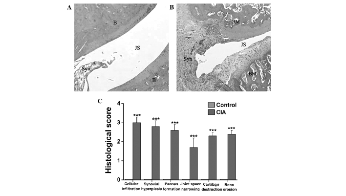
The aim of the present study was to determine a more specific, efficient and simple method for the induction of collagen-induced arthritis (CIA) in rats. Different strains of rats were injected at the base of the tail with bovine type II collagen (CII) emulsified in incomplete Freund's adjuvant (IFA). The onset and severity of arthritis were evaluated by clinical assessment. The established CIA model was analyzed using a comprehensive examination of clinical, hematological, histological and radiological parameters. The results demonstrated that Wistar rats were the most susceptible strain to CIA followed by Wistar Furth rats, with Sprague Dawley rats being the least susceptible. Following primary and booster immunization, female Wistar rats developed severe arthritis, with an incidence of >83% and low variability in clinical signs. The development of arthritis was accompanied by a significantly elevated erythrocyte sedimentation rate compared with that in the control rats. The radiographic examination revealed bone matrix resorption, considerable soft tissue swelling, periosteal new bone formation and bone erosion in the arthritic joints of the CIA rats. Histopathologically, the synovial joints of CIA rats were characterized by synovial hyperplasia, pannus formation, marked cellular infiltration, bone and cartilage erosion and narrowing of the joint space. The administration of an intradermal injection of only 200 µg bovine CII emulsified in IFA at the base of the tail therefore leads to the successful development of a CIA rat model. This well-characterized CIA rat model could be specifically used to study the pathophysiology of human rheumatoid arthritis as well as to test and develop anti-arthritic agents for humans.

Keywords: Wistar rats; bovine type II collagen; collagen-induced arthritis; incomplete Freund's adjuvant.


Figures

Figure 1.
Figure 1.
Clinical signs of CIA in the hind paws of female Wistar rats. (A and B) Representative images of the hind paws from (A) control and (B) CIA rats taken on day 15 after the primary injection of type II collagen emulsified in incomplete Freund's adjuvant. Intense edema and erythema were observed in the hind paws of the CIA rats compared with the control rats. CIA, collagen-induced arthritis.
Figure 2.
Figure 2.
Clinical progression of CIA in female Wistar rats during a 30-day course: (A) Arthritic score, (B) hind paw volume, (C) body weight and (D) ESR. Data are presented as the mean ± standard error of the mean (n=8). *P<0.05 and **P<0.01 versus control rats. CII, type II collagen; IFA, incomplete Freund's adjuvant; CIA, collagen-induced arthritis; ESR, erythrocyte sedimentation rate.
Figure 3.
Figure 3.
Radiographic evidence of CIA in the hind paws of female Wistar rats. (A and B) Representative radiographs taken on day 30 after the primary injection of type II collagen emulsified in incomplete Freund's adjuvant. Compared with the hind paws of the (A) control rats, the hind paws of the (B) CIA rats exhibited severe soft tissue swelling, bone matrix resorption, periosteal new bone formation and bone erosion. (C) Radiological analysis showed significant differences in these radiological parameters between the two groups of rats. Data are presented as the mean ± standard error of the mean (n=8). ***P<0.001 versus control rats. CIA, collagen-induced arthritis.
Figure 4.
Figure 4.
Histopathological changes in the tibiotarsal joints of female Wistar rats with CIA. (A and B) Representative hematoxylin and eosin-stained sections (magnification, ×40) revealing histopathological changes in the tibiotarsal joints of the (A) control and (B) CIA model female Wistar rats. (C) Significant differences in cellular infiltration, synovial hyperplasia, pannus formation, joint space narrowing, cartilage destruction and bone erosion in the synovial joints were found between the CIA and control rats. Data are presented as the mean ± standard error of the mean (n=8). ***P<0.001 versus control rats. CIA, collagen-induced arthritis; B, bone; JS, joint space; Syn, synovial membrane; BM, bone marrow.

Similar articles

Cited by

References

    1. Trentham DE, Townes AS, Kang AH. Autoimmunity to type II collagen: An experimental model of arthritis. J Exp Med. 1977;146:857–868. doi: 10.1084/jem.146.3.857. - DOI - PMC - PubMed
    1. Rosloniec EF, Cremer M, Kang A, Myers LK. Collagen-induced arthritis. Curr Protoc Immunol Chapter. 2001;15 doi: 10.1002/0471142735.im1505s20. Unit 15.5. - DOI - PubMed
    1. Billingham ME. Models of arthritis and the search for anti-arthritic drugs. Pharmacol Ther. 1983;21:389–428. doi: 10.1016/0163-7258(83)90062-1. - DOI - PubMed
    1. Griffiths MM. Immunogenetics of collagen-induced arthritis in rats. Int Rev Immunol. 1988;4:1–15. doi: 10.3109/08830188809044766. - DOI - PubMed
    1. Zhu L, Wei W, Zheng YQ. Effect and mechanism of action of total glucosides of paeony on synoviocytes from rats with collagen-induced arthritis. Yao Xue Xue Bao. 2006;41:166–170. (In Chinese) - PubMed