

Iris metastasis from esophageal squamous cell carcinoma: A case report
- PMID: 26622571
- PMCID: PMC4509070
- DOI: 10.3892/ol.2015.3255
Iris metastasis from esophageal squamous cell carcinoma: A case report
Abstract
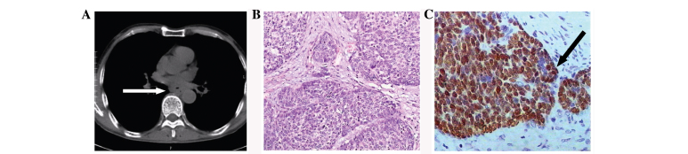
Carcinoma metastatic to the eye is a rare condition, typically associated with a poor prognosis. Breast and lung cancers are the most common sources of intraocular metastases, and the majority of metastatic lesions involve the posterior uvea, with <8% of reported cases arising in the iris. Intraocular metastasis as the presenting form of esophageal carcinoma is highly uncommon. In the present report, a rare case of metastatic iris tumor resulting from esophageal squamous cell carcinoma is discussed. A 64-year-old patient presented with a progressively distending pain in the right eye, with associated blurred vision. Local and systemic evaluation was performed, followed by treatment. Multiple examinations identified a neoplasm in the right iris and postoperative pathology revealed that the iris lesion was a metastasis of esophageal squamous cell cancer origin. The patient was treated with adjuvant radiation. To the best of our knowledge, this was only the second reported case of esophageal squamous cell carcinoma metastasizing to the iris.
Keywords: esophageal cancer; iris metastasis; squamous cell carcinoma.
Figures

Similar articles
-
Rapid progress of an iris metastasis from esophageal cancer: a case report and review of literature.Int J Ophthalmol. 2024 Aug 18;17(8):1557-1567. doi: 10.18240/ijo.2024.08.22. eCollection 2024. Int J Ophthalmol. 2024. PMID: 39156770 Free PMC article. Review.
-
Iris metastasis as the initial presentation of metastatic esophageal cancer diagnosed by fine needle aspiration biopsy: A case report.Medicine (Baltimore). 2021 Jun 4;100(22):e26232. doi: 10.1097/MD.0000000000026232. Medicine (Baltimore). 2021. PMID: 34087906 Free PMC article.
-
Snowflakes in the Eye - An Uncommon Presentation of Iris Metastasis of Esophageal Carcinoma and Review of Literature.Ocul Immunol Inflamm. 2022 Oct-Nov;30(7-8):1568-1571. doi: 10.1080/09273948.2021.1906910. Epub 2021 May 18. Ocul Immunol Inflamm. 2022. PMID: 34003704 Review.
-
[Case with metastasis of a squamous esophageal cancer of the iris with resemblance to hypopyon].Nippon Ganka Gakkai Zasshi. 2007 Sep;111(9):735-40. Nippon Ganka Gakkai Zasshi. 2007. PMID: 17907468 Japanese.
-
Small-cell lung carcinoma metastasis to the iris - case presentation.Contemp Oncol (Pozn). 2013;17(3):331-3. doi: 10.5114/wo.2013.35288. Epub 2013 Jun 28. Contemp Oncol (Pozn). 2013. PMID: 24596526 Free PMC article.
Cited by
-
Novel presentation of intraocular metastases in a patient with penile squamous cell carcinoma: a case report.J Med Case Rep. 2020 Oct 23;14(1):199. doi: 10.1186/s13256-020-02520-8. J Med Case Rep. 2020. PMID: 33092626 Free PMC article.
-
Extraocular Muscle Metastasis from Esophageal Carcinoma: an Atypical and Rare Presentation.J Gastrointest Cancer. 2018 Jun;49(2):211-213. doi: 10.1007/s12029-016-9887-4. J Gastrointest Cancer. 2018. PMID: 27785687 No abstract available.
-
Neuron-Specific Enolase and Hemoglobin as Risk Factors of Intraocular Metastasis in Patients with Renal Cell Carcinoma.Dis Markers. 2022 Apr 23;2022:2883029. doi: 10.1155/2022/2883029. eCollection 2022. Dis Markers. 2022. PMID: 35502301 Free PMC article.
-
Serum markers change for intraocular metastasis in renal cell carcinoma.Biosci Rep. 2021 Sep 30;41(9):BSR20203116. doi: 10.1042/BSR20203116. Biosci Rep. 2021. PMID: 34467977 Free PMC article.
-
Rapid progress of an iris metastasis from esophageal cancer: a case report and review of literature.Int J Ophthalmol. 2024 Aug 18;17(8):1557-1567. doi: 10.18240/ijo.2024.08.22. eCollection 2024. Int J Ophthalmol. 2024. PMID: 39156770 Free PMC article. Review.
References
-
- Parikh HK, Deshpande RK, Swaroop DV, Desai PB. Choroidal metastasis from primary adenocarcinoma of the esophagus - a case report. Indian J Cancer. 1992;29:210–214. - PubMed
LinkOut - more resources
Full Text Sources
Other Literature Sources