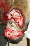
Frequency and type of toenail tumors in the dromedary camel
- PMID: 26623314
- PMCID: PMC4629581
Frequency and type of toenail tumors in the dromedary camel
Abstract
A total of 275 dromedary camels (16 males and 259 females) of local "Arabiyat" breed suffering from different types and degrees of severity of toenail tumors were surgically treated. Histopathological examination of the tissue samples removed from 50 tumor-like growths (2 males and 48 females) revealed three types of tumors; squamous cell carcinoma (70%), spiny keratoderma (22%) and fibroma (8%). An increased incidence of tumors was recorded in the medial when compared to the lateral toenails in both sexes. In females, the incidence in the medial toenails was 90/259 (34.75%) and 71/259 (27.41%) in the right and left forelimbs respectively when compared to the lateral toenails which was 25/259 (9.65%) and 5/259 (1.93%) for the respective right and left forelimbs. In the hind limbs, this ratio was 29/259 (11.20%) and 20/259 (7.72%) for right and left medial toenails respectively, whereas it was 17/259 (6.56%) and 2/259 (0.77%) for the right and left lateral toenails respectively. Similar to the observations in female camels, male camels also showed a higher incidence of these tumors in the medial when compared to the lateral toenails in both fore and hind limbs. Based on these findings, we conclude that in the dromedary camels, the medial toenails of the fore limbs are most commonly affected with tumors; with the most common tumor being the squamous cell carcinoma.
Keywords: Camels; Squamous cell carcinoma; Toenails; Tumor.
Figures


References
-
- Al-Hizab F.A, Ramadan R.O, Al-Mubarak A.I, Abdelsalam E.B. Basal cell carcinoma in a one humped camel (Camelus dromedarius). A clinical report. J. Camel Pract. Res. 2007;14(1):49–50.
-
- Gahlot T.K, Sharma C.K, Deep S, Kachhawa A.S, Sharma G.D, Chouhan D.S. Squamous cell carcinoma of the foot in a camel (Camelus dromedarius) Indian Vet. J. 1995;72(5):509–510.
-
- Goldschmidt M.H, Shofer F.S. Oxford: Butterworth Heinemann; 1998. Skin Tumors of the Dog and Cat; pp. 37–49.
-
- Janardhan K.S, Ganta C.K, Andrews G.A, Anderson D.E. Chondrosarcoma in a dromedary camel (Camelus dromedaries) J. Vet. Diag. Invest. 2011;23(3):619–622. - PubMed
-
- Murray E.F, Bravo P.W. Medicine and Surgery of Camelids. 3rd Ed. USA: Blackwell Publishing; 2010. Foot Diseases of Camelids; pp. 303–306.
LinkOut - more resources
Full Text Sources
