. 2015 Dec 1;10(12):e0143884.
doi: 10.1371/journal.pone.0143884. eCollection 2015.

Enhanced p122RhoGAP/DLC-1 Expression Can Be a Cause of Coronary Spasm

Affiliations

Enhanced p122RhoGAP/DLC-1 Expression Can Be a Cause of Coronary Spasm

Takahiko Kinjo et al. PLoS One. .

Abstract

Background: We previously showed that phospholipase C (PLC)-δ1 activity was enhanced by 3-fold in patients with coronary spastic angina (CSA). We also reported that p122Rho GTPase-activating protein/deleted in liver cancer-1 (p122RhoGAP/DLC-1) protein, which was discovered as a PLC-δ1 stimulator, was upregulated in CSA patients. We tested the hypothesis that p122RhoGAP/DLC-1 overexpression causes coronary spasm.

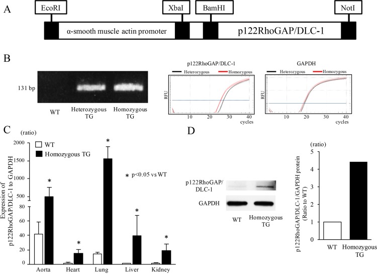
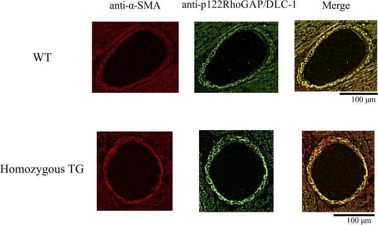
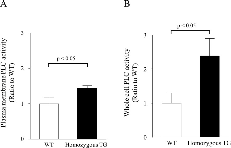
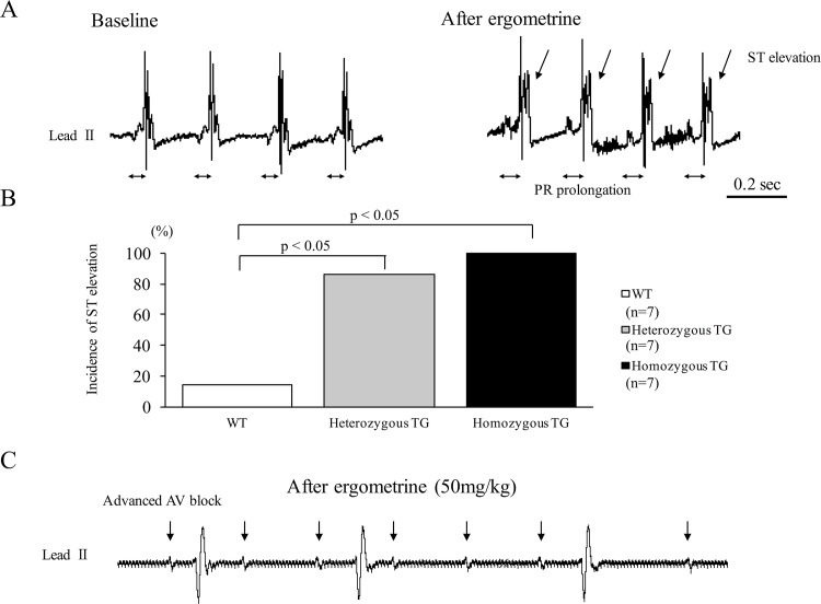
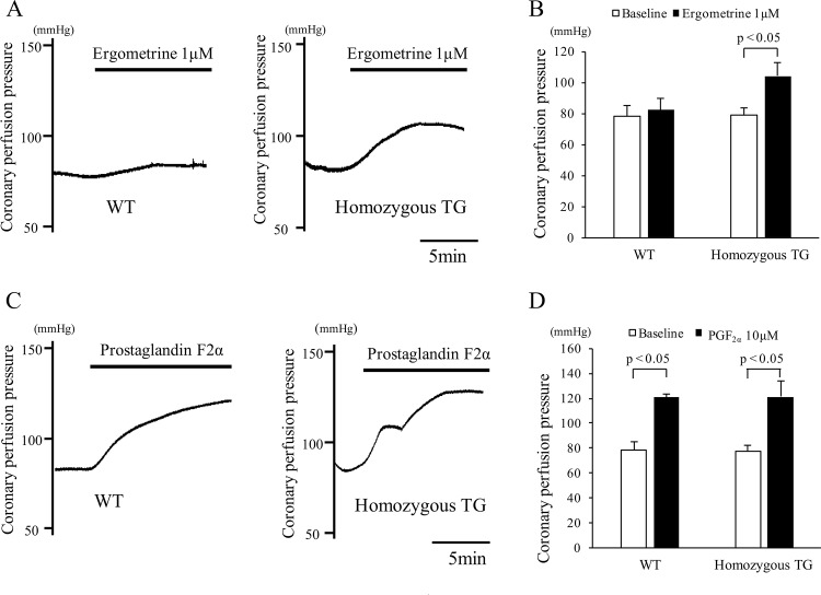
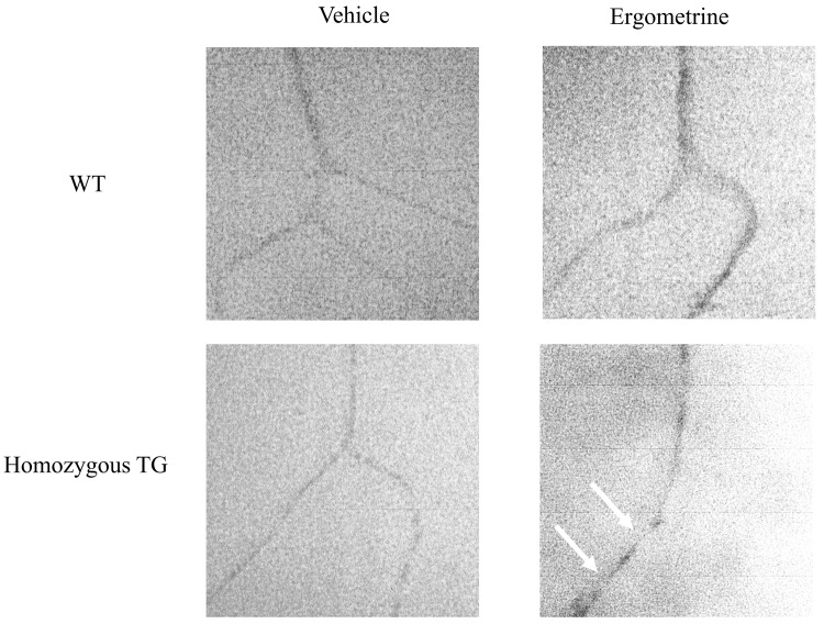
Methods and results: We generated transgenic (TG) mice with vascular smooth muscle (VSM)-specific overexpression of p122RhoGAP/DLC-1. The gene and protein expressions of p122RhoGAP/DLC-1 were markedly increased in the aorta of homozygous TG mice. Stronger staining with anti-p122RhoGAP/DLC-1 in the coronary artery was found in TG than in WT mice. PLC activities in the plasma membrane fraction and the whole cell were enhanced by 1.43 and 2.38 times, respectively, in cultured aortic vascular smooth muscle cells from homozygous TG compared with those from WT mice. Immediately after ergometrine injection, ST-segment elevation was observed in 1 of 7 WT (14%), 6 of 7 heterozygous TG (84%), and 7 of 7 homozygous TG mice (100%) (p<0.05, WT versus TGs). In the isolated Langendorff hearts, coronary perfusion pressure was increased after ergometrine in TG, but not in WT mice, despite of the similar response to prostaglandin F2α between TG and WT mice (n = 5). Focal narrowing of the coronary artery after ergometrine was documented only in TG mice.

Conclusions: VSM-specific overexpression of p122RhoGAP/DLC-1 enhanced coronary vasomotility after ergometrine injection in mice, which is relevant to human CSA.


Conflict of interest statement

Competing Interests: This study was supported in part by an Astellas/Pfizer Grant for Research on Atherosclerosis Update (http://www.jhf.or.jp/josei/12_update/) to Dr. Shibutani. There are no patents, products in development or marketed products to declare. This does not alter the authors' adherence to all the PLOS ONE policies on sharing data and materials, as detailed online in the guide for authors.

Figures

Fig 1
Fig 1. Generation of transgenic (TG) mice and their expression analyses.
(A) Schematic map of the microinjected transgene consisting of the α-smooth muscle actin promoter and the mouse p122RhoGAP/DLC-1 cDNA. (B) Representative bands (131 bp) after genomic PCR and representative amplification curves for p122RhoGAP/DLC-1 and GAPDH in real-time PCR (40 cycles). (C) The gene expression of mouse p122RhoGAP/DLC-1 in various tissues of wild type (WT) (n = 3) and homozygous TG mice (n = 3). The ratio of p122RhoGAP/DLC-1 to GAPDH expression in the liver of WT mice was used as a reference (= 1), since it showed the lowest value among the tissues of WT mice. (D) Representative bands for mouse p122RhoGAP/DLC-1 protein in the pooled aortas (left panel) and densitometric analysis between WT and homozygous TG mice (right panel). The aortas from 4 mice in WT and TG.
Fig 2
Fig 2. Immunofluorescence images of the coronary artery.
Representative images from 3 WT and 3 TG mice are shown. Left panels show anti-α-smooth muscle actin (SMA) reactivity to mark vascular smooth muscle cells (red). Middle panels show staining with an anti-p122RhoGAP/DLC-1 antibody demonstrating the increase in p122RhoGAP/DLC-1 immunoreactivity in homozygous TG mice (green). Right panels show merged images.
Fig 3
Fig 3. PLC activity in cultured vascular smooth muscle cells.
(A) PLC activity in the plasma membrane fraction (n = 3 in WT and TG mice). (B) PLC activity in the whole cell (n = 3 in WT and TG mice).
Fig 4
Fig 4. Representative ECG recordings and responses to ergometrine in wild type (WT) and transgenic (TG) mice.
(A) Representative ECG recordings before (left) and after (right) intravenous injection of ergometrine in anesthetized homozygous TG mouse. Ergometrine injection immediately elicited ST-segment elevation with PR prolongation. (B) Incidence of ST-segment elevation after ergometrine injection in WT, heterozygous, and homozygous TG mice. (C) Representative ECG showing advanced AV block in homozygous TG mice after ergometrine injection.
Fig 5
Fig 5. Coronary perfusion pressures in isolated Langendorff hearts of wild type (WT) and homozygous transgenic (TG) mice.
(A) Representative traces of coronary perfusion pressures before and after injection of ergometrine at 1 μmol/L in WT (left) and homozygous TG (right) mice. (B) Changes in coronary perfusion pressure before (open bar) and after (closed bar) injection of ergometrine at 1 μmol/L in WT (n = 5) and homozygous TG mice (n = 5). (C) Representative traces of coronary perfusion pressures before and after injection of prostaglandin F (PGF) at 10 μmol/L in WT (left) and homozygous TG (right) mice. (D) Changes in coronary perfusion pressure before (open bar) and after (closed bar) injection of PGF at 10 μmol/L in WT (n = 3) and homozygous TG mice (n = 3).
Fig 6
Fig 6. Microvascular filling experiment with microfil of the coronary arteries in wild type (WT) and homozygous transgenic (TG) mice.
In the group treated with vehicle, no focal narrowing was observed in either WT (n = 3) or TG mice (n = 3). Focal narrowing (arrows) of the coronary artery after injection of ergometrine was observed in homozygous TG (3 of 3), but not in WT mice (0 of 3).

References

    1. Oliva PB, Potts DE, Pluss RG. Coronary arterial spasm in Prinzmetal angina. Documentation by coronary arteriography. N Engl J Med. 1973;288: 745–751. - PubMed
    1. Yasue H, Touyama M, Shimamoto M, Kato H, Tanaka S. Role of autonomic nervous system in the pathogenesis of Prinzmetal's variant form of angina. Circulation. 1974;50: 534–539. - PubMed
    1. Pasupathy S, Air T, Dreyer RP, Tavella R, Beltrame JF. Systematic review of patients presenting with suspected myocardial infarction and nonobstructive coronary arteries. Circulation. 2015;131: 861–870. 10.1161/CIRCULATIONAHA.114.011201 - DOI - PubMed
    1. Kaku B, Katsuda S, Taguchi T. Life-threatening ventricular arrhythmia due to silent coronary artery spasm: usefulness of I-123 metaiodobenzylguanidine scintigraphy for detecting coronary artery spasm in the era of automated external defibrillators: a case report. J Med Case Rep. BioMed Central Ltd; 2015;9: 26 10.1186/1752-1947-9-26 - DOI - PMC - PubMed
    1. Sanna T, Lanza GA, Niccoli G, La Torre G, Cosentino N, Crea F. Coronary artery vasospasm causing ventricular fibrillation—an external loop recording. Resuscitation. 2009;80: 393–394. 10.1016/j.resuscitation.2008.12.013 - DOI - PubMed

Publication types

Substances