. 2015 Dec 1;10(12):e0143887.
doi: 10.1371/journal.pone.0143887. eCollection 2015.

Argon Mediates Anti-Apoptotic Signaling and Neuroprotection via Inhibition of Toll-Like Receptor 2 and 4

Affiliations

Argon Mediates Anti-Apoptotic Signaling and Neuroprotection via Inhibition of Toll-Like Receptor 2 and 4

Felix Ulbrich et al. PLoS One. .

Abstract

Purpose: Recently, the noble gas argon attracted significant attention due to its neuroprotective properties. However, the underlying molecular mechanism is still poorly understood. There is growing evidence that the extracellular regulated kinase 1/2 (ERK1/2) is involved in Argon´s protective effect. We hypothesized that argon mediates its protective effects via the upstream located toll-like receptors (TLRs) 2 and 4.

Methods: Apoptosis in a human neuroblastoma cell line (SH-SY5Y) was induced using rotenone. Argon treatment was performed after induction of apoptosis with different concentrations (25, 50 and 75 Vol% in oxygen 21 Vol%, carbon dioxide and nitrogen) for 2 or 4 hours respectively. Apoptosis was analyzed using flow cytometry (annexin-V (AV)/propidiumiodide (PI)) staining, caspase-3 activity and caspase cleavage. TLR density on the cells' surface was analyzed using FACS and immunohistochemistry. Inhibition of TLR signaling and extracellular regulated kinase 1/2 (ERK1/2) were assessed by western blot, activity assays and FACS analysis.

Results: Argon 75 Vol% treatment abolished rotenone-induced apoptosis. This effect was attenuated dose- and time-dependently. Argon treatment was accompanied with a significant reduction of TLR2 and TLR4 receptor density and protein expression. Moreover, argon mediated increase in ERK1/2 phosphorylation was attenuated after inhibition of TLR signaling. ERK1/2 and TLR signaling inhibitors abolished the anti-apoptotic and cytoprotective effects of argon. Immunohistochemistry results strengthened these findings.

Conclusion: These findings suggest that argon-mediated anti-apoptotic and neuroprotective effects are mediated via inhibition of TLR2 and TLR4.


Conflict of interest statement

Competing Interests: The authors have declared that no competing interests exist.

Figures

Fig 1
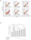
Fig 1. Argon attenuates rotenone-induced apoptosis dose-dependently but irrespective to length of treatment.
(A) Representative dot-plots of the named intervention. (B) Annexin V positive and PI negative cells [%] of rotenone-induced apoptosis and consecutive argon treatment (n = 8; mean±SD; untreated 11.3±3.3% vs. rotenone 24.1±1.8% and rotenone 24.1±1.8% vs. argon 75 Vol% 14.3±1.8% and 50 Vol% 16.8±2.0% at 4 hours and rotenone 24.1±1.8% vs. argon 75 Vol% 14.8±2.4% and 50 Vol% 17.4±2.4% at 2 hours; all *** = p<0.001 and rotenone 24.1±1.8% vs. argon 25 Vol% 19.1±2.7% at 4 hours and rotenone 24.1±1.8% vs. argon 25 Vol% 21.0±2.7% at 2 hours; both ** = p<0.01).
Fig 2
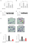
Fig 2. Argon reduces TLR2 and TLR4 receptor density.
(A and B) FACS analysis of TLR2 and TLR4 receptor density on the cells´ surface (n = 8; mean±SD; rotenone 2.87±0.49% vs. rotenone+argon 75 Vol% [2 h] 2.070.4±3; * = p<0.05; rotenone 3.0±20.34 vs. rotenone+argon Vol%75 [2 h] 2.41±0.34; ** = p<0.01 and untreated was set to 1 vs. rotenone 2.87±0.49% and 3.02±0.46%; both *** = p<0.001). TLR2 and TLR4 protein expression. (C and D) Representing Western blot images, representing TLR2 (C) and TLR4 (D) protein expression after treatment with rotenone and argon (75 Vol%, 2 h). TLR2 and TLR4 expression following argon treatment. (E) Representative histological images of TLR2 (brown color) and TLR4 (red color) in differently treated cells (yellow arrow: TLR2 expression after rotenone treatment, green arrow: TLR4 expression after rotenone treatment). (F) Analysis of histogram quantification, left panel: TLR2 histogram (n = 5; TLR2 untreated 12±9; argon 75 Vol% 52±12; rotenone 205±41 vs. rotenone+argon 75 Vol% [2 h] 63±28; *** = p<0.001) and right panel: TLR4 histogram (n = 5; TLR untreated; 52±17, argon 75 Vol% 61±19; rotenone 168±32 vs. rotenone+argon 75 Vol% [2 h] 103±27; *** = p<0.01).
Fig 3
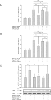
Fig 3. Argon-mediated cytoprotective effects are abolished after TLR2 and TLR4 inhibition.
Annexin V positive and PI negative cells [%] of rotenone-induced apoptosis and consecutive argon treatment (n = 8; mean±SD; rotenone 19.4±0.7 vs. rotenone+argon 75 Vol% [2 h] 13.1±1.4; *** = p<0.001), (A) with TLR2 inhibitor (TAK-242; rotenone+argon 75 Vol% [2 h] 13.1±1.4 vs. TAK-242+rotenone+argon 75 Vol% [2 h] 16.9±1.0; ** = p<0.01 and rotenone 19.4±0.7 vs. TAK-242+rotenone+argon 75 Vol% [2 h] 16.9±1.0 and TAK-242+rotenone 18.5±1.6 vs. TAK-242+rotenone+argon 75 Vol% [2 h] 16.9±1.0; both * = p<0.05) or (B) TLR2 and TLR4 inhibitor (OxPAPC; rotenone+argon 75 Vol% [2 h] 13.1±1.4 vs. OxPAPC+rotenone+argon 75 Vol% [2 h] 18.1±2.9; *** = p<0.001, rotenone 19.4±0.7 vs. OxPAPC+rotenone+argon 75 Vol% [2 h] 18.1±2.9 and OxPAPC+rotenone 18.6±1.5 vs. OxPAPC+rotenone+argon 75 Vol% [2 h] 18.1±2.9; both n.s. = not significant). Argon-mediated phosphorylation of IRAK4 is abolished after TLR2 and TLR4 inhibition. (C) Densitometric analysis and representative Western blot image of IRAK4 protein phosphorylation after rotenone treatment. Equal loading was confirmed by reprobing with total-IRAK4 and analysis was performed in relation to the corresponding protein (n = 8; mean±SD; untreated 0.70±0.13 vs. rotenone 1.21±0.19; *** = p<0.001; rotenone 1.21±0.19 vs. rotenone+argon 75 Vol% [2 h] 0.80±0.13; ** = p<0.01 and rotenone+argon 75 Vol% [2 h] 0.80±0.13 vs. OxPAPC+rotenone+argon 75 Vol% [2 h] 0.92±0.2; * = p<0.05).
Fig 4
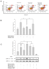
Fig 4. Argon´s cytoprotective properties are mediated via the phosphorylation of ERK1/2, which directly depends on TLR2 and TLR4 signalling.
(A) Representative dot-plots of the particular intervention. (B) Annexin V positive and PI negative cells [%] of rotenone-induced apoptosis, consecutive argon treatment and inhibition of ERK1/2 using FR180204 (n = 8; mean±SD; rotenone 11.3±3.3% vs. rotenone+argon 75 Vol% [2 h] 13.1±2.6% and rotenone+argon 75 Vol% [2 h] 13.1±2.6% vs. FR180204+rotenone+argon 75 Vol% [2 h] 21.2±1.9%; both *** = p<0.001) (C) Densitometric analysis and representative Western blot image, demonstrating the phosphorylation of ERK1/2 protein expression after treatment with rotenone, argon and inhibitors of ERK, TLR2 and TLR4. Equal loading was confirmed by reprobing with the non-phosphorylated protein form (t-ERK; n = 8; mean±SD; untreated 0.31±0.07 vs. rotenone 0.66±0.11, rotenone 0.66±0.11 vs. argon 75 Vol% [2 h] 0.42±0.06 and rotenone 0.66±0.11 vs. rotenone+argon 75 Vol% [2 h] 0.93±0.10; all *** = p<0.001; rotenone+argon 75 Vol% [2 h] 0.93±0.10 vs. OxPAPC+rotenone+argon 75 Vol% [2 h] 0.66±0.09; ** = p<0.01 and rotenone+argon 75 Vol% [2 h] 0.93±0.10 vs. TAK-242+rotenone+argon 75 Vol% [2 h] 0.75±0.12, * = p<0.05).
Fig 5
Fig 5. Argon-mediated decrease of apoptosis is attenuated following ERK1/2 and TLR inhibition.
(A) Caspase activity assay demonstrating argon´s effect on enzyme activity in the context of different inhibitors (n = 8, mean±SD; untreated 369±60 vs. rotenone 2458±149; rotenone 2458±149 vs. rotenone+argon 75 Vol% [2 h] 956±163, rotenone+argon 75 Vol% [2 h] 956±163 vs. OxPAPC+rotenone+argon 75 Vol% [2 h] 1789±160, rotenone+argon 75 Vol% [2 h] 956±163 vs. TAK-242+rotenone+argon 75 Vol% [2 h] 1641±182 and rotenone+argon 75 Vol% [2 h] 956±163 vs. FR180204+rotenone+argon 75 Vol% [2 h] 2062±149; all *** = p<0.001). (B) Diagram depicting the proposed mechanism of argon-mediated protective effects on neuronal cells. Argon treatment affects both, TLR2 and TLR4, attenuates IRAK4 phosphorylation but not MyD88 and subsequently increases ERK1/2 phosphorylation in this model of rotenone-induced apoptosis. As a consequence, caspase-3 cleavage and activity are reduced thus conferring cytoprotection. Inhibition of TLRs and ERK1/2 MAPK abolished argon mediated effects regarding cytoprotection.

References

    1. Lopez AD, Mathers CD, Ezzati M, Jamison DT, Murray CJ. Global and regional burden of disease and risk factors, 2001: systematic analysis of population health data. Lancet. 2006;367(9524):1747–57. - PubMed
    1. Frost RB, Farrer TJ, Primosch M, Hedges DW. Prevalence of traumatic brain injury in the general adult population: a meta-analysis. Neuroepidemiology. 2013;40(3):154–9. 10.1159/000343275 - DOI - PubMed
    1. Langlois JA, Rutland-Brown W, Wald MM. The epidemiology and impact of traumatic brain injury: a brief overview. The Journal of head trauma rehabilitation. 2006;21(5):375–8. - PubMed
    1. Tymianski M. Novel approaches to neuroprotection trials in acute ischemic stroke. Stroke; a journal of cerebral circulation. 2013;44(10):2942–50. 10.1161/STROKEAHA.113.000731 - DOI - PubMed
    1. Ulbrich F, Schallner N, Coburn M, Loop T, Lagreze WA, Biermann J, et al. Argon inhalation attenuates retinal apoptosis after ischemia/reperfusion injury in a time- and dose-dependent manner in rats. PloS one. 2014;9(12):e115984 10.1371/journal.pone.0115984 - DOI - PMC - PubMed

Publication types

MeSH terms