. 2016 Jan 5;7(1):845-59.
doi: 10.18632/oncotarget.6417.

Synergistic cooperation between ABT-263 and MEK1/2 inhibitor: effect on apoptosis and proliferation of acute myeloid leukemia cells

Affiliations

Synergistic cooperation between ABT-263 and MEK1/2 inhibitor: effect on apoptosis and proliferation of acute myeloid leukemia cells

Kelly Airiau et al. Oncotarget. .

Abstract

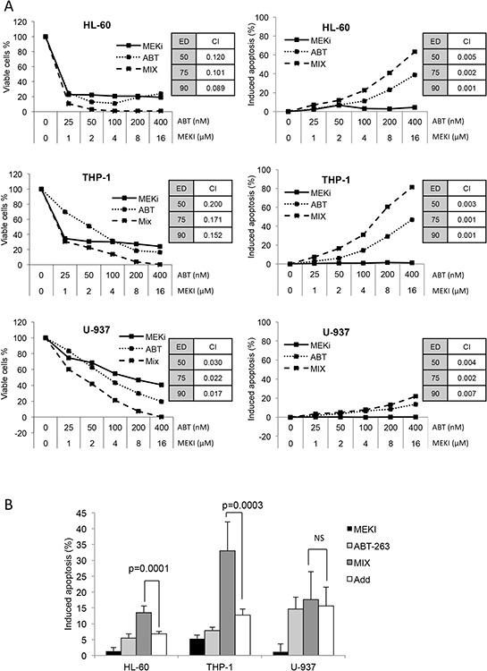
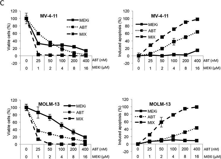
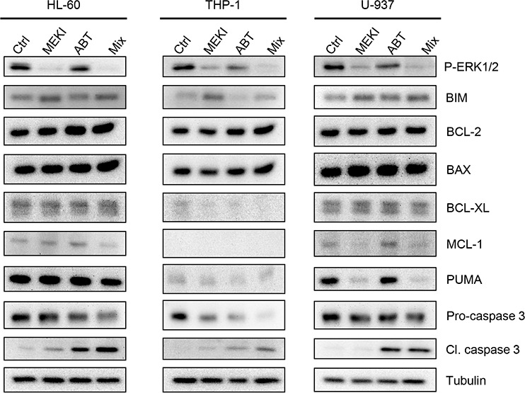
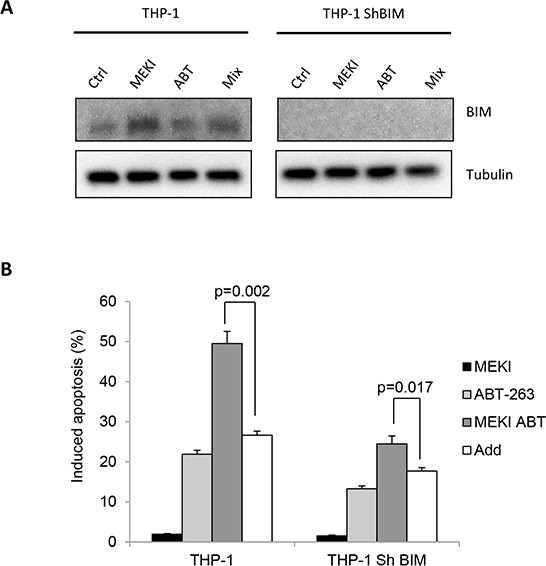
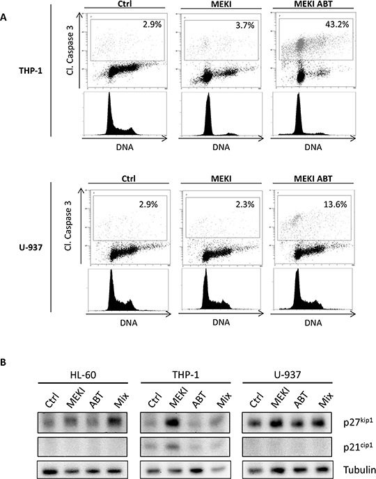
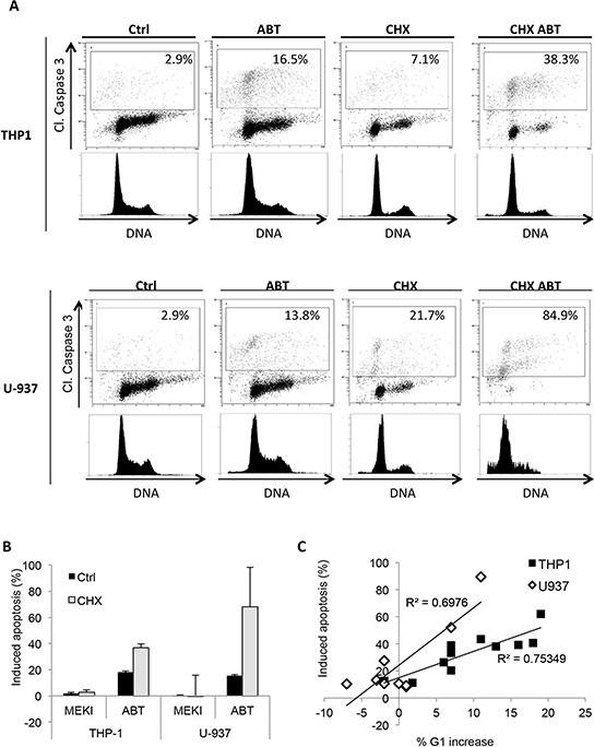
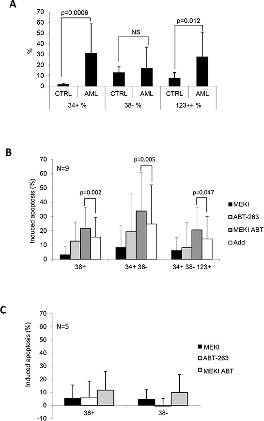
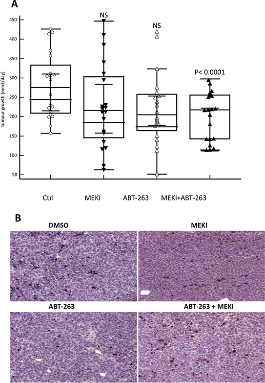
In spite of intensive research to improve treatment of acute myeloid leukemia (AML) more than half of all patients continue to develop a refractory disease. Therefore there is need to improve AML treatment. The overexpression of the BCL-2 family anti-apoptotic members, like BCL-2 or BCL-xL has been largely reported in lymphoid tumors but also in AML and other tumors. To counteract the anti-apoptotic effect of BCL-2, BH3 mimetics have been developed to target cancer cells. An increase in activity of ERK1/2 mitogen activated protein (MAP) kinase has also been reported in AML and might be targeted by MEK1/2 inhibitors. Hence, in the current work, we investigated whether the association of a BH3 mimetic such ABT-263 and the MEK1/2 inhibitor pimasertib (MEKI), was efficient to target AML cells. A synergistic increasing of apoptosis was observed in AML cell lines and in primary cells without affecting normal bone marrow cells. Such cooperation was confirmed on tumor growth in a mouse xenograft model of AML. In addition we demonstrated that MEKI sensitized the cells to apoptosis through its ability to promote a G1 cell cycle arrest. So, this combination of a MAP Kinase pathway inhibitor and a BH3 mimetic could be a promising strategy to improve the treatment of AML.

Keywords: BH3 mimetic drugs; MEK inhibitors; acute myeloid leukemia.


Conflict of interest statement

CONFLICTS OF INTEREST

The authors declare no conflict of interest.

Figures

Figure 1
Figure 1. MEKI and ABT-263 synergize to inhibit cell proliferation and induce apoptosis in AML cell lines
A. and C. HL-60, THP-1 or U-937 (A) and MV-4–11 or MOLM-13 (C) cells were incubated with increasing concentrations of ABT-263, MEKI or both at a constant ratio (Mix). The amount of viable cells was measured through their ATP content by chemoluminescence after a 72-h culture (left column). The percentage of apoptotic cells was determined by flow cytometry with a MMP probe after a 24-h incubation (right column). Combination indexes were calculated at ED 50, 75 and 90 using the Calcusyn software. B. HL-60, THP-1 or U-937 cells were treated for 24 h with 1 μM MEKI (black bars), 200 nM ABT-263 (light grey bars) or both (MIX, grey bars). Apoptosis was then measured by flow cytometry as described in Figure 1. Mean +/− SD of seven experiments is shown. The calculated additive effect of both drugs is also shown (white bars) and compared to the measured MIX effect using the Student paired t test. C. MV-4–11 or MOLM-13 (C) cells were incubated with increasing concentrations of ABT-263, MEKI or both at a constant ratio (Mix). The amount of viable cells was measured through their ATP content by chemoluminescence after a 72-h culture (left column). The percentage of apoptotic cells was determined by flow cytometry with a MMP probe after a 24-h incubation (right column).
Figure 2
Figure 2. Protein expression of the Bcl-2 family members following ABT-263 and MEKI treatment
HL-60, THP-1 or U-937 cells were analyzed by western blot for expression of the Bcl-2 family proteins. The three cell lines were treated for 24 h with 1 μM MEKI, 200 nM ABT-263 or both (Mix) and analyzed for Erk1/2 phosphorylation, BIM, Bcl-2, BAX, Bcl-XL, Mcl-1 and PUMA expression and Caspase 3 cleavage.
Figure 3
Figure 3. BIM accumulation does not participate in the cooperation between ABT-263 and MEKI to induce apoptosis in THP-1
THP-1 cells or THP-1 cells depleted for BIM by RNA interference were treated with 1 μM MEKI, 200 nM ABT-263 or both in combination (Mix) for 24 h and analyzed for BIM expression A. and apoptosis induction B.
Figure 4
Figure 4. ABT-263-induced apoptosis depends on the cell cycle block in G1 phase
A. THP-1 and U-937 cells were untreated or treated with 1 μM MEKI or a combination of 1 μM MEKI and 200 nM ABT-263 for 24 h. Cells were then fixed, permeabilized and stained with anti-cleaved caspase 3 antibody (Y axis) and Vibrant Violet Cycle probe (X axis). Cells were then analyzed by flow cytometry. B. HL-60, THP-1 or U-937 cells were treated for 24 h with 1 μM MEKI, 200 nM ABT-263 or both (Mix) and analyzed by western blot for p27kip1, p21cip1 and tubulin expression.
Figure 5
Figure 5. CHX induces a cell cycle block in G1 phase and synergizes with ABT-263 to increase apoptosis
A. THP-1 and U-937 cells were pre-treated or not with 3 μM CHX for 24 h and treated or not with 200 nM ABT-263 for 24 h. Cells were then fixed, permeabilized and stained with anti-cleaved caspase 3 antibody (Y axis) and Vibrant Violet Cycle probe (X axis) and analyzed by flow cytometry. B. THP-1 and U-937 cells were pre-treated or not with 3 μM CHX for 24 h and treated or not with 1 μM MEKI, 200 nM ABT-263 or both (Mix) for 24 h. Cells were then fixed, permeabilized and stained with anti-cleaved caspase 3 antibody and analysed by flow cytometry. Mean +/− SD of three experiments is shown. C. THP-1 (black squares) and U-937 (white diamonds) cells were pre-treated with MEKI (200 nM) or CHX (3 μM) for 24 h. Samples were then treated or not with 200 nM ABT-263 for 24 h and analyzed by flow cytometry for cell cycle and caspase 3 activation. The graph represents the correlation between the percentage of ABT-263-induced apoptosis and the increased amount of cells in G1 phase.
Figure 6
Figure 6. MEKI and ABT-263 synergize to induce apoptosis in leukemic stem cells
Mononuclear cells from AML or normal control bone marrows were labelled with APC-CD34, PC5-CD38, PE-CD123 and FITC-annexin V. A. The percentage of CD34+ cells, CD38- cells in the CD34+ population and CD123+ cells in the CD34+/CD38- population was plotted for normal control and AML samples. Results are expressed as mean +/–SD and are representative of 10 and 17 experiments for respectively control and AML bone marrows. P indicates the significance using the Mann-Whitney test. Annexin-V+ cells were considered as apoptotic and their percentage was evaluated in the CD34+/CD38+, CD34+/CD38-, and CD34+/CD38-/CD123+ populations. B. Apoptosis induction was evaluated in 9 AML samples as indicated in each of the three populations after treatment with MEKI (black bars), ABT-263 (white bars) or both (grey bars). C: Apoptosis induction was evaluated in 6 normal control samples in the CD34+/CD38+ and CD34+/CD38- populations.
Figure 7
Figure 7. MEKI and ABT-263 synergize to reduce tumor growth in vivo
A. HL60 cells were subcutaneously injected in NSG mice and mice were treated five days a week with vehicle control, MEKI (0.2 mg/kg), ABT-263 (1 mg/kg) or both. The growth rate was calculated from the measurement of the tumour volume twice a week and defined as the increase in mm3/day in each mouse. The graph represents the values of two independent experiments performed on 10 mice/group for each experiment. B. Histological sections of tumours stained with anti-activated caspase 3 antibody. A significant field of slides in tumours from either vehicle-, MEKI-, ABT-263- or MEKI + ABT-263-fed mice is shown as indicated.

References

    1. Shipley JL, Butera JN. Acute myelogenous leukemia. Exp Hematol. 2009;37:649–58. - PubMed
    1. Juliusson G, Antunovic P, Derolf A, Lehmann S, Mollgard L, Stockelberg D, et al. Age and acute myeloid leukemia: real world data on decision to treat and outcomes from the Swedish Acute Leukemia Registry. Blood. 2009;113:4179–87. - PubMed
    1. Miesner M, Haferlach C, Bacher U, Weiss T, Macijewski K, Kohlmann A, et al. Multilineage dysplasia (MLD) in acute myeloid leukemia (AML) correlates with MDS-related cytogenetic abnormalities and a prior history of MDS or MDS/MPN but has no independent prognostic relevance: a comparison of 408 cases classified as “AML not otherwise specified” (AML-NOS) or “AML with myelodysplasia-related changes” (AML-MRC) Blood. 2010;116:2742–51. - PubMed
    1. Leith CP, Kopecky KJ, Godwin J, McConnell T, Slovak ML, Chen IM, et al. Acute myeloid leukemia in the elderly: assessment of multidrug resistance (MDR1) and cytogenetics distinguishes biologic subgroups with remarkably distinct responses to standard chemotherapy. A Southwest Oncology Group study. Blood. 1997;89:3323–9. - PubMed
    1. Estey E. Acute myeloid leukemia and myelodysplastic syndromes in older patients. J Clin Oncol. 2007;25:1908–15. - PubMed

Publication types

MeSH terms