. 2016 Feb 9;23(2):350-9.
doi: 10.1016/j.cmet.2015.10.018. Epub 2015 Nov 25.

Pdgfrβ+ Mural Preadipocytes Contribute to Adipocyte Hyperplasia Induced by High-Fat-Diet Feeding and Prolonged Cold Exposure in Adult Mice

Affiliations

Pdgfrβ+ Mural Preadipocytes Contribute to Adipocyte Hyperplasia Induced by High-Fat-Diet Feeding and Prolonged Cold Exposure in Adult Mice

Lavanya Vishvanath et al. Cell Metab. .

Abstract

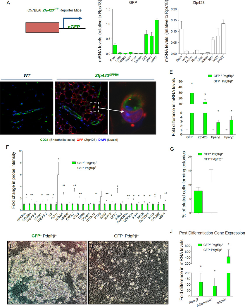
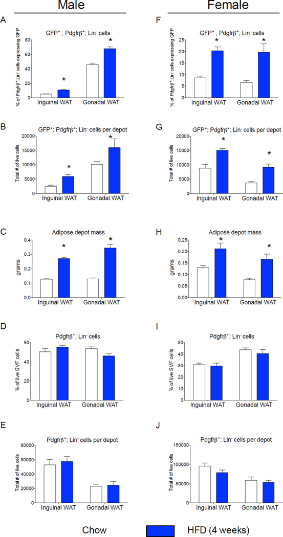
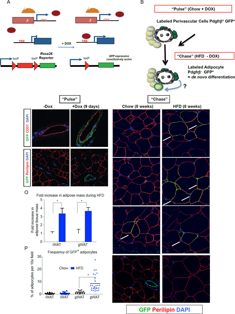
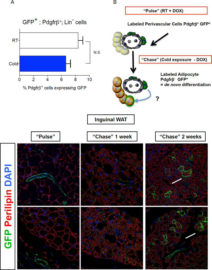
The expansion of white adipose tissue (WAT) in obesity involves de novo differentiation of new adipocytes; however, the cellular origin of these cells remains unclear. Here, we utilize Zfp423(GFP) reporter mice to characterize adipose mural (Pdgfrβ(+)) cells with varying levels of the preadipocyte commitment factor Zfp423. We find that adipose tissue contains distinct mural populations, with levels of Zfp423 distinguishing adipogenic from inflammatory-like mural cells. Using our "MuralChaser" lineage tracking system, we uncover adipose perivascular cells as developmental precursors of adipocytes formed in obesity, with adipogenesis and precursor abundance regulated in a depot-dependent manner. Interestingly, Pdgfrβ(+) cells do not significantly contribute to the initial cold-induced recruitment of beige adipocytes in WAT; it is only after prolonged cold exposure that these cells differentiate into beige adipocytes. These results provide genetic evidence for a mural cell origin of white adipocytes in obesity and suggest that beige adipogenesis may originate from multiple sources.


Conflict of interest statement

Conflicting interests statement. The authors declare that they have no competing financial interests.

Figures

Figure 1
Figure 1. Zfp423-expressing adipose mural cells are lineage-primed adipose precursors
(A) mRNA levels of GFP and endogenous Zfp423 in different adult tissues isolated from newly derived C57BL/6 Zfp423GFPB6 reporter mice in which enhanced GFP (eGFP) is expressed under the control of a Zfp423-containing BAC. (B–D) Confocal images of cryosectioned inguinal white adipose tissue (WAT) stained with antibodies recognizing GFP (red) and the endothelial protein CD31 (green), with nuclei counterstained with DAPI. (B) WAT from wild-type (WT) mice. (C) WAT from Zfp423GFPB6 mice. (D) Digital enhancement of the small blood vessel shown in (C) (white arrow). Note expression of GFP in mature adipocytes and subset of periendothelial cells. Similar results were observed in gonadal WAT (E). Quantitative PCR measurements of mRNA levels of GFP, Zfp423, and Pparγ isoforms in freshly isolated GFP+ or GFP mural (Pdgfrβ+; Lin) cells from the gonadal WAT of male Zfp423GFPB6 mice. * denotes p <0.05 from student’s t-test. Bars represent mean ± SEM. n= 3 samples, with each sample representing cells obtained from pooled tissue from 6–7 male mice. (F) Relative probe intensity values of key inflammatory genes with an overlapping presence in multiple gene sets enriched in GFP mural cells. Bars represent mean ± SEM. * and ** denotes p <0.05 and p<0.005, respectively (G) Quantification of colony-forming unit potential of freshly isolated GFP+ or GFP mural cells. n=8 (H–I) Phase contrast images of spontaneous differentiation in cultures of GFP+ (H) and GFP (I) mural cells maintained at confluence in growth media containing 2% FBS and 1% ITS (insulin, transferrin, and selenium). Images captured 10 days post-confluence. (J) mRNA levels of adipocyte selective genes in differentiated cultures from (H–I) n=3 Bars represent mean ± SEM. * and ** denotes p <0.05.
Figure 2
Figure 2. The frequency of Zfp423-expressing adipose mural cells is regulated in a sex- and depot-dependent manner
(A) Percentage of Pdgfrβ+;Lin cells expressing GFP (GFP+; Pdgfrβ+; Lin) in WAT depots of Zfp423GFPB6 male mice fed chow or high fat diet (HFD) for 4 weeks following weaning. (B) Total number of GFP+; Pdgfrβ+; Lin cells per individual WAT depot after 4 weeks of chow or HFD. (C) Adipose depot mass (grams) after 4 weeks of chow or HFD. (D) Percentage of the live cell population of the total SVF that are Pdgfrβ+;Lin cells. (E) Total number of live Pdgfrβ+;Lin cells per individual WAT depot after 4 weeks of chow or HFD. (F–J) Corresponding data in female animals fed chow or HFD for the same duration. * denotes p <0.05 from student’s t-test. Bars represent mean ± SEM. n= 5–6 animals for each group.
Figure 3
Figure 3. Lineage tracing reveals Pdgfrβ+ cells as a cellular origin of adipogenesis associated with high-fat diet feeding
(A) Genetic alleles comprising of the “MuralChaser” lineage tracing system. In the presence of doxycycline (DOX) rtTA activates Cre expression. Cre excises the loxP-flanked membrane tdTomato cassette and allows constitutive activation of membrane GFP (mGFP) reporter expression. (B) “Pulse-Chase” lineage tracing approach. DOX-containing food (600 mg/kg) is administered to male MuralChaser mice for 9 days to label Pdgfrβ+ cells (“Pulse”). Animals are then immediately harvested (Pulse) or are switched to a high-fat diet (HFD) or chow diet for 8 weeks in the absence of DOX (Chase). mGFP+ adipocytes represent de novo differentiated fat cells formed during the 8 week period. (C–D) mGFP labeling is confined to peri-endothelial (CD31+) cells and occurs in a DOX-dependent manner. (E–F) mGFP labeling is not found in mature adipocytes (perilipin+). (G–I) Following 8 weeks of chow feeding, only few mGFP+ gonadl adipocytes (gWAT) are observed. (J–L) Following 8 weeks of HFD feeding, numerous mGFP+ perilipin+ adipocytes are observed. These cells represent de novo differentiated adipocytes formed during the 8 week period. (M–N) After 8 weeks of chow or HFD, very few mGFP+ adipocytes are observed in the inguinal WAT (iWAT), supporting the notion of very little postnatal adipocyte turnover or hyperplasia in this depot during HFD feeding. Note that antibody staining was utilized with formalin-fixed paraffin embedded sections; native mGFP and mTdTomato fluorescence are not observed here and are lost during tissue processing. (O) Fold increase in adipose tissue mass (normalized to body weight) during 8-week HFD period. Bars represent mean + stdev. * in both panels denotes p < 0.05 from student’s t-test. (P) Quantification of mGFP+ adipocytes observed in randomly chosen 10× magnification fields from gWAT and iWAT sections. * denotes p <0.05 from student’s t-test. Twenty-six random 10x magnification fields were photographed from duplicate sections of stained WAT depots from four animals of each group. All images show were photographed at 63× magnification.
Figure 4
Figure 4. Pdgfrβ+ mural cells contribute to beige adipocyte formation in adult WAT after prolonged cold exposure
(A) Percentage of Pdgfrβ+;Lin cells expressing GFP (GFP+; Pdgfrβ+; Lin) in WAT depots of Zfp423GFPB6 male mice housed at room temperature or cold exposed (6°C) for 2 weeks. Bars represent mean ± SEM. n= 5–6 animals for each group. (B) “Pulse-Chase” lineage tracing approach. DOX-containing food (600 mg/kg) is administered to MuralChaser mice for 9 days at room temperature to label Pdgfrβ+ cells (“Pulse”). Afterwards, animals were immediately harvested (Pulse) or are housed in the cold (6 °C) for either 1 or 2 weeks in the absence of DOX (Chase). mGFP+ multilocular adipocytes represent de novo differentiated fat cells formed during the cold exposure period. (C–D) Confocal immunofluorescence image of inguinal WAT from MuralChaser mice dox-treated for 9 days. Parrafin–embedded sections are immunostained with antibodies recognizing mGFP and perilipin. mGFP staining is confined to the vasculature at this stage. (E–F) Confocal immunofluorescence image of inguinal WAT from MuralChaser mice after 1 week of cold exposure (after removal of DOX). mGFP staining is still largely confined to the vasculature at this stage. (G–H) Confocal immunofluorescence image of inguinal WAT from MuralChaser mice after 2 weeks of cold exposure (after removal of DOX). Several small clusters of mGFP+ multilocular adipocytes can now be observed, indicating a contribution of Pdgfrβ+ mural cells to beige adipogenesis during prolonged cold exposure. All images photographed at 63× magnification.

References

    1. Armulik A, Genove G, Betsholtz C. Pericytes: developmental, physiological, and pathological perspectives, problems, and promises. Dev Cell. 2011;21:193–215. - PubMed
    1. Berry R, Jeffery E, Rodeheffer MS. Weighing in on adipocyte precursors. Cell Metab. 2014;19:8–20. - PMC - PubMed
    1. Berry R, Rodeheffer MS. Characterization of the adipocyte cellular lineage in vivo. Nat Cell Biol. 2013;15:302–308. - PMC - PubMed
    1. Cawthorn WP, Scheller EL, MacDougald OA. Adipose tissue stem cells meet preadipocyte commitment: going back to the future. J Lipid Res. 2012;53:227–246. - PMC - PubMed
    1. Chau YY, Bandiera R, Serrels A, Martinez-Estrada OM, Qing W, Lee M, Slight J, Thornburn A, Berry R, McHaffie S, et al. Visceral and subcutaneous fat have different origins and evidence supports a mesothelial source. Nat Cell Biol. 2014;16:367–375. - PMC - PubMed

Publication types

MeSH terms