MRI thresholds for discrimination between normal and mild temporomandibular joint involvement in juvenile idiopathic arthritis
- PMID: 26626730
- PMCID: PMC4665947
- DOI: 10.1186/s12969-015-0051-7
MRI thresholds for discrimination between normal and mild temporomandibular joint involvement in juvenile idiopathic arthritis
Abstract
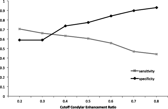
Background: Currently there is no consensus agreement on the degree of enhancement in normal temporomandibular joints (TMJ) in children, which makes it difficult for clinicians to distinguish between the presence/absence of mild synovitis. Quantitative measurements of synovial and condylar enhancement may be useful additions to current qualitative methods on early MRI diagnosis and follow up of TMJ involvement in JIA. The purpose of the study is to establish thresholds/tendencies for quantitative measures that enable distinction between mild TMJ involvement and normal TMJ appearance based on the degree of synovial and bone marrow enhancement in JIA patients.
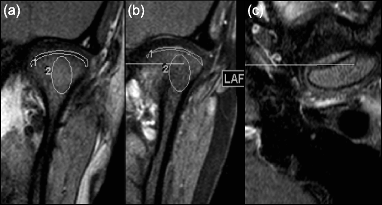
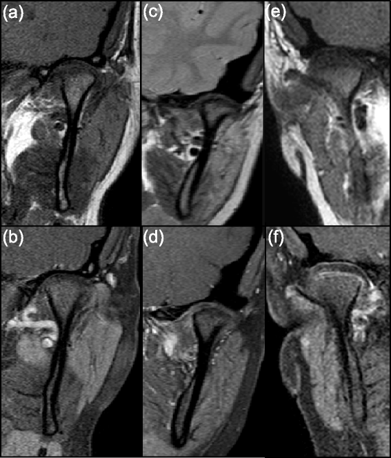
Methods: TMJ MRI examinations in 67 children with JIA and in 24 non-rheumatologic children who underwent MRI for neurologic/orbit indications were retrospectively assessed. As a priori determined TMJs of JIA patients were categorized into three groups by experienced staff radiologists based on the degree of synovial and condylar enhancement: no active disease (rheumatologic control), mild and moderate/severe findings. The signal intensity (SI) of the synovial tissue around each condyle and of the bone marrow was measured to calculate the enhancement ratio (ER) and relative SI change. The ER was calculated using signal to noise ratios, while relative SI change was calculated using signal intensities alone. Quantitative measurements of synovial and condylar enhancement of TMJs with mild or moderate/severe findings were compared with the rheumatologic and non-rheumatologic controls.
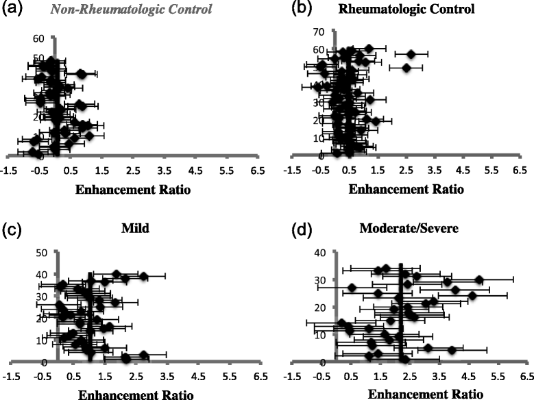
Results: Mean ER values were significantly different between the TMJs without active disease and those with mild and moderate/severe synovial enhancement, with highest values in the moderate/severe group (P < 0.0001). Similar findings were seen for condylar enhancement with P < 0.005. Relative SI change was unable to differentiate TMJs with mild synovitis from the two controls (P > 0.10). 27/60 (45%) TMJs without active disease had osteochondral changes. 8/40 (20%) TMJs in the mild group did not demonstrate any synovial thickening.
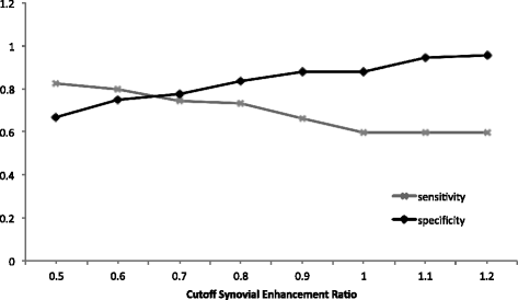
Conclusions: Quantitative signal to noise ratios of TMJ synovial and condylar enhancement generate thresholds/tendencies, which offer additional information to differentiate mild synovitis from normal TMJs in JIA patients. Osteochondral changes and synovial thickening may not be reliable indicators of active TMJ involvement and should be differentiated from synovial enhancement.
Figures


References
-
- Kuseler A, Pedersen TK, Gelineck J, Herlin T. A 2 year followup study of enhanced magnetic resonance imaging and clinical examination of the temporomandibular joint in children with juvenile idiopathic arthritis. J Rheumatol. 2005;32(1):162–9. - PubMed
-
- Weiss PF, Arabshahi B, Johnson A, Bilaniuk LT, Zarnow D, Cahill AM, et al. High prevalence of temporomandibular joint arthritis at disease onset in children with juvenile idiopathic arthritis, as detected by magnetic resonance imaging but not by ultrasound. Arthritis Rheum. 2008;58(4):1189–96. doi: 10.1002/art.23401. - DOI - PubMed
-
- Sarnat B. The temporomandibular joint. Springfield, Illinois: Charles C. Thomas; 1951.
MeSH terms
LinkOut - more resources
Full Text Sources
Other Literature Sources
Medical
Research Materials

