

Cell Death Signaling
- PMID: 26626938
- PMCID: PMC4665079
- DOI: 10.1101/cshperspect.a006080
Cell Death Signaling
Abstract
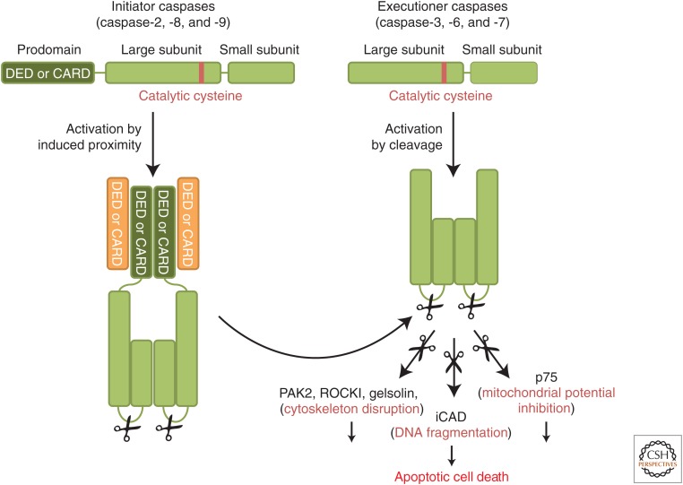
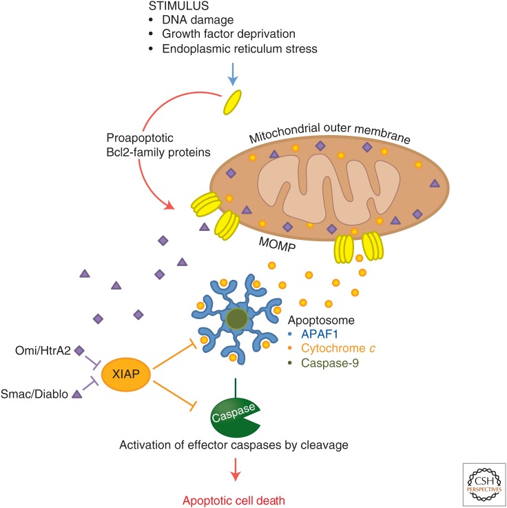
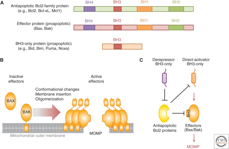
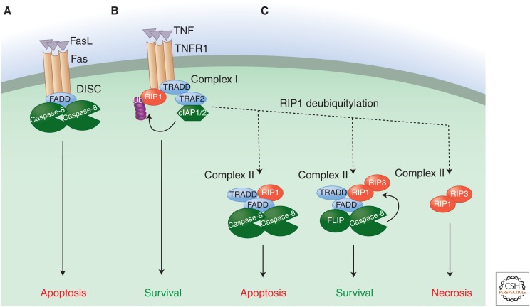
In multicellular organisms, cell death is a critical and active process that maintains tissue homeostasis and eliminates potentially harmful cells. There are three major types of morphologically distinct cell death: apoptosis (type I cell death), autophagic cell death (type II), and necrosis (type III). All three can be executed through distinct, and sometimes overlapping, signaling pathways that are engaged in response to specific stimuli. Apoptosis is triggered when cell-surface death receptors such as Fas are bound by their ligands (the extrinsic pathway) or when Bcl2-family proapoptotic proteins cause the permeabilization of the mitochondrial outer membrane (the intrinsic pathway). Both pathways converge on the activation of the caspase protease family, which is ultimately responsible for the dismantling of the cell. Autophagy defines a catabolic process in which parts of the cytosol and specific organelles are engulfed by a double-membrane structure, known as the autophagosome, and eventually degraded. Autophagy is mostly a survival mechanism; nevertheless, there are a few examples of autophagic cell death in which components of the autophagic signaling pathway actively promote cell death. Necrotic cell death is characterized by the rapid loss of plasma membrane integrity. This form of cell death can result from active signaling pathways, the best characterized of which is dependent on the activity of the protein kinase RIP3.
Copyright © 2015 Cold Spring Harbor Laboratory Press; all rights reserved.
Figures
References
-
- Allan LA, Clarke PR. 2007. Phosphorylation of caspase-9 by CDK1/cyclin B1 protects mitotic cells against apoptosis. Mol Cell 26: 301–310. - PubMed
-
- Allan LA, Clarke PR. 2009. Apoptosis and autophagy: Regulation of caspase 9 by phosphorylation. FEBS J 276: 6063–6073. - PubMed
-
- Allan LA, Morrice N, Brady S, Magee G, Pathak S, Clarke PR. 2003. Inhibition of caspase-9 through phosphorylation at Thr 125 by ERK MAPK. Nat Cell Biol 5: 647–654. - PubMed
-
- Alves NL, Derks IA, Berk E, Spijker R, van Lier RA, Eldering E. 2006. The Noxa/Mcl-1 axis regulates susceptibility to apoptosis under glucose limitation in dividing T cells. Immunity 24: 703–716. - PubMed
Publication types
MeSH terms
Substances
Grants and funding
LinkOut - more resources
Full Text Sources
Other Literature Sources
Medical
Research Materials
Miscellaneous