

Oxidative stress in multiple sclerosis: Central and peripheral mode of action
- PMID: 26626971
- PMCID: PMC7094520
- DOI: 10.1016/j.expneurol.2015.11.010
Oxidative stress in multiple sclerosis: Central and peripheral mode of action
Abstract
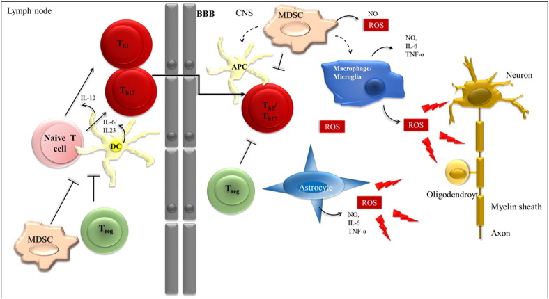
Accumulating evidence suggests that oxidative stress plays a major role in the pathogenesis of multiple sclerosis (MS). Reactive oxygen species (ROS), which if produced in excess lead to oxidative stress, have been implicated as mediators of demyelination and axonal damage in both MS and its animal models. One of the most studied cell populations in the context of ROS-mediated tissue damage in MS are macrophages and their CNS companion, microglia cells. However, and this aspect is less well appreciated, the extracellular and intracellular redox milieu is integral to many processes underlying T cell activation, proliferation and apoptosis. In this review article we discuss how oxidative stress affects central as well as peripheral aspects of MS and how manipulation of ROS pathways can potentially affect the course of the disease. It is our strong belief that the well-directed shaping of ROS pathways has the potential to ameliorate disease progression in MS.
Accumulating evidence suggests that oxidative stress plays a major role in the pathogenesis of multiple sclerosis (MS). Reactive oxygen species (ROS), which if produced in excess lead to oxidative stress, have been implicated as mediators of demyelination and axonal damage in both MS and its animal models. One of the most studied cell populations in the context of ROS-mediated tissue damage in MS are macrophages and their CNS companion, microglia cells. However, and this aspect is less well appreciated, the extracellular and intracellular redox milieu is integral to many processes underlying T cell activation, proliferation and apoptosis. In this review article we discuss how oxidative stress affects central as well as peripheral aspects of MS and how manipulation of ROS pathways can potentially affect the course of the disease. It is our strong belief that the well-directed shaping of ROS pathways has the potential to ameliorate disease progression in MS.
- •
Reactive oxygen species play a major role in the pathogenesis of multiple sclerosis and contribute to demyelination in the CNS.
- •
Redox states influence T cells, which are activated in the periphery and are strongly associated with MS pathogenesis.
- •
We summarize central and peripheral mode of actions of ROS in MS.
- •
We discuss treatment options, which target oxidative stress pathways, with regard to central and peripheral effects.
Keywords: DMF; Neurodegeneration; Neuroprotection; Nrf2.
Figures


References
-
- Altmeyer P.J., Matthes U., Pawlak F., Hoffmann K., Frosch P.J., Ruppert P., Wassilew S.W., Horn T., Kreysel H.W., Lutz G. Antipsoriatic effect of fumaric acid derivatives. Results of a multicenter double-blind study in 100 patients. J. Am. Acad. Dermatol. 1994;30:977–981. - PubMed
-
- Angelini G., Gardella S., Ardy M., Ciriolo M.R., Filomeni G., Di Trapani G., Clarke F., Sitia R., Rubartelli A. Antigen-presenting dendritic cells provide the reducing extracellular microenvironment required for T lymphocyte activation. Proc. Natl. Acad. Sci. U. S. A. 2002;99:1491–1496. - PMC - PubMed
-
- Barnett M.H., Prineas J.W. Relapsing and remitting multiple sclerosis: pathology of the newly forming lesion. Ann. Neurol. 2004;55:458–468. - PubMed
-
- Bo L., Geurts J.J., Mork S.J., van der Valk P. Grey matter pathology in multiple sclerosis. Acta Neurol. Scand. Suppl. 2006;183:48–50. - PubMed
Publication types
MeSH terms
LinkOut - more resources
Full Text Sources
Other Literature Sources
Medical
