. 2016 Jan 22;291(4):2004-2017.
doi: 10.1074/jbc.M115.685628. Epub 2015 Dec 1.

Non-native Conformers of Cystic Fibrosis Transmembrane Conductance Regulator NBD1 Are Recognized by Hsp27 and Conjugated to SUMO-2 for Degradation

Affiliations

Non-native Conformers of Cystic Fibrosis Transmembrane Conductance Regulator NBD1 Are Recognized by Hsp27 and Conjugated to SUMO-2 for Degradation

Xiaoyan Gong et al. J Biol Chem. .

Abstract

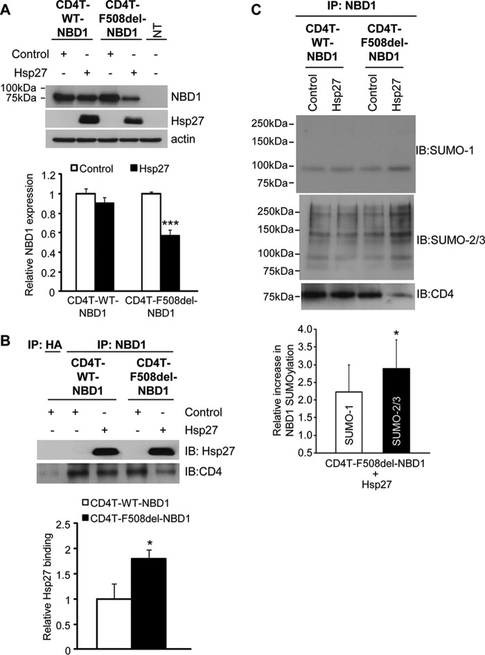
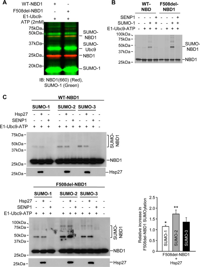
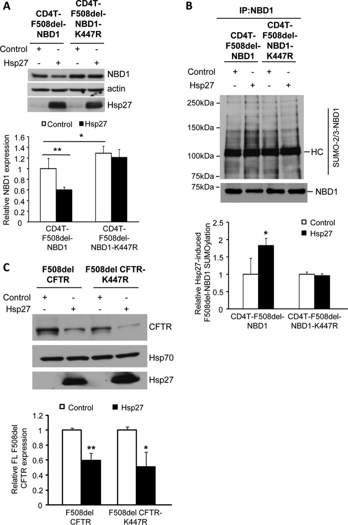
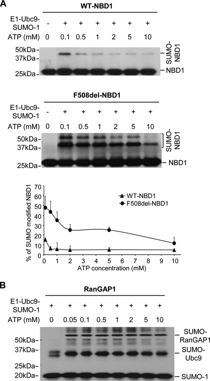
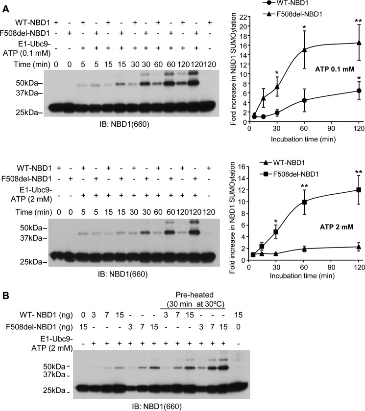
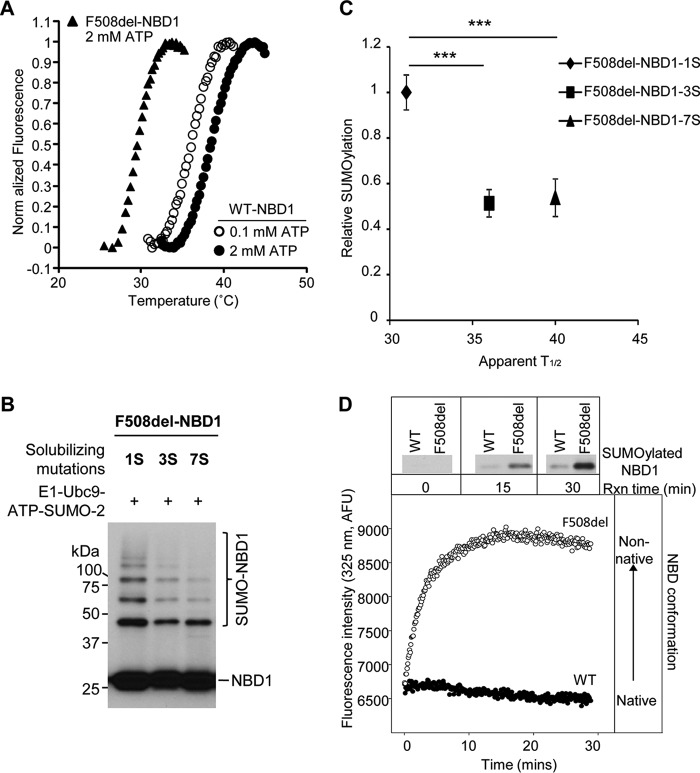
A newly identified pathway for selective degradation of the common mutant of the cystic fibrosis transmembrane conductance regulator (CFTR), F508del, is initiated by binding of the small heat shock protein, Hsp27. Hsp27 collaborates with Ubc9, the E2 enzyme for protein SUMOylation, to selectively degrade F508del CFTR via the SUMO-targeted ubiquitin E3 ligase, RNF4 (RING finger protein 4) (1). Here, we ask what properties of CFTR are sensed by the Hsp27-Ubc9 pathway by examining the ability of NBD1 (locus of the F508del mutation) to mimic the disposal of full-length (FL) CFTR. Similar to FL CFTR, F508del NBD1 expression was reduced 50-60% by Hsp27; it interacted preferentially with the mutant and was modified primarily by SUMO-2. Mutation of the consensus SUMOylation site, Lys(447), obviated Hsp27-mediated F508del NBD1 SUMOylation and degradation. As for FL CFTR and NBD1 in vivo, SUMO modification using purified components in vitro was greater for F508del NBD1 versus WT and for the SUMO-2 paralog. Several findings indicated that Hsp27-Ubc9 targets the SUMOylation of a transitional, non-native conformation of F508del NBD1: (a) its modification decreased as [ATP] increased, reflecting stabilization of the nucleotide-binding domain by ligand binding; (b) a temperature-induced increase in intrinsic fluorescence, which reflects formation of a transitional NBD1 conformation, was followed by its SUMO modification; and (c) introduction of solubilizing or revertant mutations to stabilize F508del NBD1 reduced its SUMO modification. These findings indicate that the Hsp27-Ubc9 pathway recognizes a non-native conformation of mutant NBD1, which leads to its SUMO-2 conjugation and degradation by the ubiquitin-proteasome system.

Keywords: ABC transporter; Prot; SUMO; SUMOylation; chloride channel; cystic fibrosis; cystic fibrosis transmembrane conductance regulator (CFTR); post-translational modification (PTM); protein conformation; protein degradation.


Figures

FIGURE 1.
FIGURE 1.
Selective Hsp27 impact on F508del NBD1 expression, binding, and SUMOylation in vivo. A, overexpression of Hsp27 promotes the degradation of CD4T-F508del-NBD1. HEK293 cells were transiently transfected with CD4T-WT-NBD1 or CD4T-F508del-NBD1 and with vector control or Hsp27 as described under “Experimental Procedures.” Whole cell lysates were extracted 48 h after transfection, and protein expression was detected by IB using the indicated antibodies. NBD1 protein levels were quantified and normalized to control values from four independent experiments. ***, p = 0.0003. B, Hsp27 preferentially interacts with CD4T-F508del-NBD1. CD4T-WT-NBD1 or CD4T-F508del-NBD1 was expressed in HEK293 cells as in A. The interaction of the NBDs with Hsp27 was evaluated by IP using anti-NBD1 followed by resolution of the co-IPed proteins by SDS-PAGE and IB with antibodies against Hsp27 or CD4. The densities of the Hsp27 blots were normalized to the amount of NBD1 in the IP. The quantitative data normalized to WT binding level are the averages of three independent experiments. *, p = 0.032. C, Hsp27 promotes NBD1 SUMOylation in vivo. CD4T-WT-NBD1 or CD4T-F508del-NBD1 and vector control or Hsp27 were expressed as above. After 48 h, the cells were lysed, and co-IP was performed using anti-NBD1 and resolved on SDS-PAGE and IB with antibodies against SUMO paralogs. The endogenous SUMO signals were normalized to the amount of NBD1 pulled down (CD4 IB) and then compared with their respective control values in three independent experiments. *, p = 0.023.
FIGURE 2.
FIGURE 2.
In vitro SUMOylation and dependence on Hsp27. A, identification of SUMO-1 modified WT and F508del NBD1. Immunoblots were performed using monoclonal anti-NBD1 (antibody 660) and polyclonal anti-SUMO-1 antibodies, detected with IRDye 680RD donkey anti-mouse or IRDye 800CW donkey anti-rabbit secondary antibodies using a Li-Cor Odyssey. B, selective SUMO-1 modification of F508del-NBD1 requires the SUMO E1 and E2-conjugating enzymes and ATP and is reversed by the SUMO protease, SENP-1. Purified 1S-NBD1, WT or F508del, was incubated with SUMOylation components for 60 min at 27 °C. The reaction mixture was resolved by SDS-PAGE, and NBD1 was detected by antibody (antibody 660). C, preferential modification of F508del-NBD1 by SUMO-2 in vitro is augmented by Hsp27. SUMOylation reactions were run with purified paralogs, and NBD1 was detected as in B. SUMOylated F508del-NBD1 densities were quantified and normalized to their respective control values from three independent experiments. *, p = 0.023; **, p = 0.008.
FIGURE 3.
FIGURE 3.
Hsp27-mediated degradation and SUMOylation of F508del-NBD1 in vivo requires Lys447. A, the SUMO consensus site, Lys447, was mutated to generate CD4T-F508del-NBD1-K447R; this mutant and its control were expressed in HEK293 cells with Hsp27 or vector control, as above. After 48 h, F508del-NBD1 expression was assessed by IB using the indicated antibodies. Protein levels were quantified and normalized to the vector control from three independent experiments. **, p = 0.001; *, p < 0.05. B, HEK293 cells were transfected with the plasmids used in A. NBD1 SUMOylation was detected by NBD1 IP followed by IB with antibodies against NBD1 or endogenous SUMO-2/3. HC, antibody heavy chain. The SUMO signal was normalized to the amount of NBD1 pulled down and then normalized to control; data from three independent experiments. *, p = 0.007. C, K447R does not abrogate the impact of Hsp27 on FL F508del degradation. F508del CFTR or F508del CFTR-K447R and Hsp27 or control plasmids were transfected into HEK293 cells as above. Protein expression was detected by IB using the indicated antibodies. F508del CFTR levels in Hsp27 co-expressing cells were normalized to their respective controls from four independent experiments. **, p = 0.008.
FIGURE 4.
FIGURE 4.
Structural analysis of the location of Lys447 using experimental x-ray structures of NBD1. A and B, the experimentally determined structure of NBD1 is shown as a ribbon (A) or the calculated electrostatic surface (B). The Lys447 residue is shown as a red sphere in the ribbon, and its contribution to the surface on NBD1 is circled in the surface model. The location of the Phe508 residue is shown as magenta spheres for reference in A. The images were rendered using Protein Data Bank code 2BBO for NBD1 (35). As noted in the text, surface exposure of Lys447 is also predicted in the full-length homology model of Serohijos et al. (34).
FIGURE 5.
FIGURE 5.
Dependence of WT- and F508del-NBD1 SUMOylation on ATP concentration in vitro. A, ATP concentration was varied as indicated under otherwise standard in vitro assay conditions. The graph plots the percentages of total NBD1 signal detected at higher molecular mass, as indicated; the data are from three independent experiments. B, ATP concentration dependence of in vitro SUMOylation of the model substrate, RanGAP1, run under the standard assay conditions and detected by SUMO-1 IB.
FIGURE 6.
FIGURE 6.
SUMOylation kinetics and [ATP] dependence. These data were used to establish the standard assay conditions and for subsequent experiments relating to NBD1 conformation. A, time courses of WT and F508del NBD1 modification by SUMO-1 at 0.1 or 2 mm ATP at 27 °C. Immunoblots were performed using NBD1 (antibody 660) antibody. Quantitation of the data is provided to the right, where density values were normalized to values at 5 min; the mean data are derived from three independent experiments. B, in vitro SUMOylation of WT or F508del NBD1 at three levels of purified protein, run for 1 h at 27 °C. The indicated reactions included preheating to 30 °C for 30 min prior to adding the SUMOylation reagents for the standard assay. Controls were performed without added E1, E2, or ATP (left and right lanes).
FIGURE 7.
FIGURE 7.
SUMOylation is inversely proportional to NBD1 thermal stability. A, temperature-dependent unfolding of NBD1. Purified WT and F508del 1S-NBDs were diluted into buffers, and the fluorescence of SYPRO orange, which interacts with exposed hydrophobic regions, was monitored. The apparent T½ values for the unfolding transitions of WT NBD1 at 2 mm ATP (closed circles) and 0.1 mm ATP (open circles) and for F508del NBD1 at 2 mm ATP (triangles) are provided in the text and in C and are in agreement with previously determined values (25). B, solubilizing and revertant mutations (25) reduce the extent of F508del NBD1 SUMOylation in vitro. C, relation between relative in vitro SUMOylation (Fig. 7B) and thermal stability data for 1S, 3S, and 7S F508del NBD1s (25) (***, p = 0.0003, 3S; p = 0.0002, 7S). D, SUMOylation follows the kinetics of NBD1 transition to a non-native conformation. WT and F508del NBD1 SUMO modification (upper panels) were determined at 0.1 mm ATP and 27 °C at the times indicated (data from Fig. 6A). Immunoblots were performed using NBD1 (660) antibody. Intrinsic fluorescence values at 325 nm were normalized to the values at 5 min for WT and F508del 1S-NBD1.

References

    1. Ahner A., Gong X., Schmidt B. Z., Peters K. W., Rabeh W. M., Thibodeau P. H., Lukacs G. L., and Frizzell R. A. (2013) Small heat shock proteins target mutant cystic fibrosis transmembrane conductance regulator for degradation via a small ubiquitin-like modifier-dependent pathway. Mol. Biol. Cell 24, 74–84 - PMC - PubMed
    1. Frizzell R. A., and Hanrahan J. W. (2012) Physiology of epithelial chloride and fluid secretion. Cold Spring Harb. Perspect. Med. 2, a009563. - PMC - PubMed
    1. Farinha C. M., and Amaral M. D. (2005) Most F508del-CFTR is targeted to degradation at an early folding checkpoint and independently of calnexin. Mol. Cell Biol. 25, 5242–5252 - PMC - PubMed
    1. Rowe S. M., and Verkman A. S. (2013) Cystic fibrosis transmembrane regulator correctors and potentiators. Cold Spring Harb. Perspect. Med. 3, a009761. - PMC - PubMed
    1. Clancy J. P., Rowe S. M., Accurso F. J., Aitken M. L., Amin R. S., Ashlock M. A., Ballmann M., Boyle M. P., Bronsveld I., Campbell P. W., De Boeck K., Donaldson S. H., Dorkin H. L., Dunitz J. M., Durie P. R., Jain M., Leonard A., McCoy K. S., Moss R. B., Pilewski J. M., Rosenbluth D. B., Rubenstein R. C., Schechter M. S., Botfield M., Ordoñez C. L., Spencer-Green G. T., Vernillet L., Wisseh S., Yen K., and Konstan M. W. (2012) Results of a phase IIa study of VX-809, an investigational CFTR corrector compound, in subjects with cystic fibrosis homozygous for the F508del-CFTR mutation. Thorax 67, 12–18 - PMC - PubMed

Publication types

MeSH terms

Substances

Associated data