. 2015 Dec 1;13(9):1781-8.
doi: 10.1016/j.celrep.2015.10.067. Epub 2015 Nov 25.

The Binding Interface between Human APOBEC3F and HIV-1 Vif Elucidated by Genetic and Computational Approaches

Affiliations

The Binding Interface between Human APOBEC3F and HIV-1 Vif Elucidated by Genetic and Computational Approaches

Christopher Richards et al. Cell Rep. .

Abstract

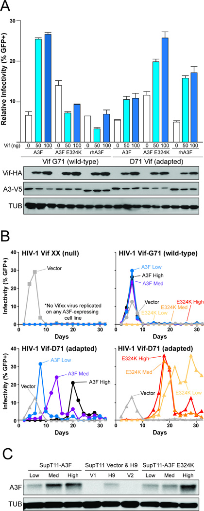
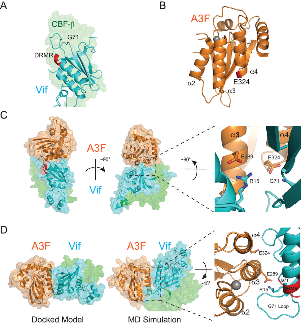
APOBEC3 family DNA cytosine deaminases provide overlapping defenses against pathogen infections. However, most viruses have elaborate evasion mechanisms such as the HIV-1 Vif protein, which subverts cellular CBF-β and a polyubiquitin ligase complex to neutralize these enzymes. Despite advances in APOBEC3 and Vif biology, a full understanding of this direct host-pathogen conflict has been elusive. We combine virus adaptation and computational studies to interrogate the APOBEC3F-Vif interface and build a robust structural model. A recurring compensatory amino acid substitution from adaptation experiments provided an initial docking constraint, and microsecond molecular dynamic simulations optimized interface contacts. Virus infectivity experiments validated a long-lasting electrostatic interaction between APOBEC3F E289 and HIV-1 Vif R15. Taken together with mutagenesis results, we propose a wobble model to explain how HIV-1 Vif has evolved to bind different APOBEC3 enzymes and, more generally, how pathogens may evolve to escape innate host defenses.

Keywords: APOBEC3F; APOBEC3F-Vif interface; HIV-1; Vif; pathogen-host interaction.


Figures

Figure 1
Figure 1. HIV-1 Adaptation to Vif-Resistant A3F
(A) A schematic depicting the co-culture selection strategy used to adapt HIV-1 to Vif-resistant A3F expressing cells as the selective pressure. Viruses are passaged step-wise approximately every 8–11 days from permissive to increasingly non-permissive cultures as shown. Upon completing a round of selection from fully permissive to fully non-permissive cultures, portions of each culture were cycled back to the beginning of the process for another round of selection. (B) Anti-A3F immunoblot of SupT11-derived T cell lines stably expressing human A3F, A3F QE323-324EK, or empty vector (relative protein expression levels: L, low; M, medium; H, high; VH, very high; V, empty vector). A3F levels in HIV-1 infected primary T-cell and H9 lysates are shown for comparison. (C) Bar graphs indicating the number of independent times that each of the indicated HIV-1 IIIB or LAI Vif amino acid changes were observed. The x-axis is a to-scale depiction of Vif residues 1–192 with previously reported motifs indicated below for reference. In most instances, HIV-1IIIB and HIV-1LAI had the same amino acid change at a given position with the exception of HIV-1IIIB E117K and HIV-1LAI D117N for both A3F QE323-324EK and rhA3F selective conditions (shown explicitly in the figure).
Figure 2
Figure 2. HIV-1 Vif G71D Enables Viral Infectivity in the Presence of Vif-Resistant A3F
(A) Single-cycle infectivity data for Vif-null HIV-1IIIB produced in the presence of huA3F, huA3F E324K, or rhA3F and the indicated amounts of Vif G71 (wild-type) or Vif D71 (adapted) expression constructs. Immunoblots are shown below for Vif (anti-HA), A3F (anti-V5), and tubulin (anti-TUB). (B) Spreading infection data for HIV-1IIIB stocks with the indicated Vif alleles in SupT11 clones expressing zero (empty vector), low, medium, or high levels of huA3F or huA3F E324K. (C) Anti-A3F immunoblot of SupT11-derived T cell lines stably expressing huA3F or A3F E324K. Empty vector-transfected SupT11 clones V1 and V2 and the non-permissive T cell line H9 are shown for comparison.
Figure 3
Figure 3. Vif-A3F Interaction Model
(A) A ribbon schematic of HIV-1 Vif highlighting residues D14, R15, M16, R17, and G71. D14 is the first residue of an α-helix containing Vif residues 14–31, which includes the DRMR motif. Vif G71 is located in a nearby loop on the same surface of the structure (PDB: 4N9F). Vif is colored cyan, and a faded surface representation of CBF-β is shown in green to facilitate visualization. (B) A ribbon schematic of the Vif-binding domain of huA3F highlighting the α3 and α4 helices and the position of residue E324 near the end of the α4-helix (PDB: 4IOU). (C) A model of the complex generated by docking A3Fctd onto Vif. In this initial docked model, direct interactions occur between A3F E324 and Vif G71 as well as A3F E289 and R15. (D) An MD simulation-optimized model of the A3F-Vif macromolecular complex. Residues E289 and R15 form a strong persistent interaction, and residues within the Vif G71 containing loop are interacting with A3F residues between helices α2 and α3. See the main text for details.
Figure 4
Figure 4. HIV-1 Vif-A3F Interface Interactions Validated by Gain-of-Function Viral Infectivity Experiments
(A) Single-cycle infectivity data for Vif-null HIV-1IIIB produced in the presence of huA3F, huA3F E324K, or huA3F E289K and the indicated amounts of Vif R15 (wild-type) or Vif E15 expression constructs. Each histogram bar shows the average infectivity from three independent experiments (mean ± SEM). Statistical significance was determined by two-way ANOVA followed by a post hoc Bonferroni test to determine significance between or within testing groups. P values of less than 0.05 are considered significant. Immunoblots are shown below for virus-like particles (anti-A3F and anti-P24) and producer cell lysates (anti-A3F, anti-Vif, and anti-TUB). (B) Spreading infection data demonstrating that HIV-1 encoding Vif R15E replicates on SupT11 that stably expresses A3F E289K, whereas HIV-1 IIIB Vif cannot establish a productive infection in identical culture conditions. (C) SupT11-derived T cell lines used for these spreading infections have been stably transfected with human A3F, A3F E289K, or a vector. A3F protein expression in H9 lysates is shown for comparison.
Figure 5
Figure 5. Wobble Model to Explain the Evolution of the Vif-A3 Interaction
The schematic shows the wobble model for adaptation of the Vif side of the Vif-A3 interface. Each hexagon of the lattice depicts a potential interaction: ancestral, dark gray; potential, light blue; attenuated, light gray; A3F, orange; A3G, purple. The model predicts that the ancestral Vif-A3 interaction was strong and consisted of six interaction points (arbitrary number for discussion purposes). Viral transmission to a new host with a larger A3 repertoire results in diminished but still partly functional interactions. A series of rapid adaptations (possibly most or all in the original new host) restores the strong interaction and results in partly overlapping interaction surfaces. Then, over a much longer evolutionary period, the combined effect of many independent wobbles triggered by virus or host genetic changes could result in the present day non-overlapping surfaces of Vif that interact with A3F and A3G. Similar rules would apply to other HIV-1 restrictive A3s (not shown for simplicity). A strong interaction may be disrupted by a viral or host amino acid substitution mutation, but the overall interaction can be restored by a compensatory change in Vif at the same site, nearby, or even to extend the edge of an interaction surface. If this change occurs at a new position, it can be considered a ‘wobble’. A series of wobbles over evolutionary time can account for a shift in the entire interaction surface.

References

    1. Albin JS, Hache G, Hultquist JF, Brown WL, Harris RS. Long-term restriction by APOBEC3F selects human immunodeficiency virus type 1 variants with restored Vif function. J Virol. 2010a;84:10209–10219. - PMC - PubMed
    1. Albin JS, LaRue RS, Weaver JA, Brown WL, Shindo K, Harjes E, Matsuo H, Harris RS. A single amino acid in human APOBEC3F alters susceptibility to HIV-1 Vif. J Biol Chem. 2010b;285:40785–40792. - PMC - PubMed
    1. Anderson BD, Harris RS. Transcriptional regulation of APOBEC3 antiviral immunity through the CBF-β/RUNX axis. Science Advances. 2015 in press. - PMC - PubMed
    1. Bishop KN, Holmes RK, Sheehy AM, Davidson NO, Cho SJ, Malim MH. Cytidine deamination of retroviral DNA by diverse APOBEC proteins. Curr Biol. 2004;14:1392–1396. - PubMed
    1. Bohn MF, Shandilya SM, Albin JS, Kouno T, Anderson BD, McDougle RM, Carpenter MA, Rathore A, Evans L, Davis AN, et al. Crystal structure of the DNA cytosine deaminase APOBEC3F: the catalytically active and HIV-1 Vif-binding domain. Structure. 2013;21:1042–1050. - PMC - PubMed

Publication types

MeSH terms

Substances

LinkOut - more resources