. 2016 Feb 9:314:35-46.
doi: 10.1016/j.neuroscience.2015.11.048. Epub 2015 Nov 26.

Role of estradiol in intrinsic hindbrain AMPK regulation of hypothalamic AMPK, metabolic neuropeptide, and norepinephrine activity and food intake in the female rat

Affiliations

Role of estradiol in intrinsic hindbrain AMPK regulation of hypothalamic AMPK, metabolic neuropeptide, and norepinephrine activity and food intake in the female rat

F S H Alenazi et al. Neuroscience. .

Abstract

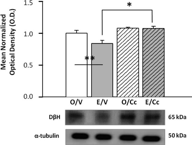
This study addressed the hypothesis that dorsomedial hindbrain adenosine 5'-monophosphate-activated protein kinase (AMPK) imposes inherent estradiol-dependent control of hypothalamic AMPK, neuropeptide, and norepinephrine (NE) activity and feeding in the female rat. Estradiol (E)- or oil (O)-implanted ovariectomized rats were injected with the AMPK inhibitor compound c (Cc) or vehicle into the caudal fourth ventricle (CV4) prior to micropunch-dissection of individual hypothalamic metabolic loci or assessment of food intake. Cc decreased hindbrain dorsal vagal complex phosphoAMPK (pAMPK) in both E and O; tissue ATP levels were reduced by this treatment in O only. In E/Cc, pAMPK expression was diminished in the lateral hypothalamic area (LHA) and ventromedial (VMH) and paraventricular (PVH) nuclei; only PVH pAMPK was suppressed by this treatment in O/Cc. Cc decreased PVH corticotropin-releasing hormone and arcuate (ARH) proopiomelanocortin (POMC) and neuropeptide Y in O, but suppressed only POMC in E. O/Cc exhibited both augmented (PVH, VMH) and decreased (LHA, ARH) hypothalamic NE content, whereas Cc treatment of E elevated preoptic and dorsomedial hypothalamic nucleus NE. Cc completely or incompletely repressed feeding in E versus O, respectively. Results implicate dorsomedial hindbrain AMPK in physiological stimulus-induced feeding in females. Excepting POMC, hypothalamic neuropeptide responses to this sensor may be contingent on estrogen. Estradiol likely designates hypothalamic targets of altered NE signaling due to hindbrain AMPK activation. Divergent changes in NE content of hypothalamic loci in O/Cc uniquely demonstrate sensor-induced bimodal catecholamine signaling to those sites.

Keywords: ATP; adenosine 5′-monophosphate-activated protein kinase; compound C; estradiol; norepinephrine; pro-opiomelanocortin.


Figures

Figure 1
Figure 1. Effects of Caudal Fourth Ventricular (CV4) Administration of the Adenosine 5’-Monophosphate-Activated Protein Kinase (AMPK) Inhibitor, Compound C (Cc), on Hindbrain Caudal Dorsal Vagal Complex (cDVC) AMPK and phosphoAMPK (pAMPK) Levels in Estradiol (E)- and Oil (O)-Implanted Ovariectomized (OVX) Female Rats
Groups of E and O animals were sacrificed 1 hr after Cc administration (n=5 O/Cc, n=5 E/Cc) or V (n=5 O/V, n=5 E/V). Bars in Figures 1.A and 1.B depict mean corresponding normalized cDVC AMPK and pAMPK protein optical density (O.D.) measures ± S.E.M. for O/V [solid white bar], E/V [solid grey bar], O/Cc [diagonal-striped white bar], and E/Cc [diagonal-striped gray bar] treatment groups. Representative immunoblots of AMPK, pAMPK, and the housekeeping protein α-tubulin are shown below respective graphs. cDVC pAMPK data analysis revealed significant main (F3,12=53.9; p<0.0001), Cc (F=125.9; p<0.0001), and E (F=33.7; p<0.0001) effects. Bars in Figure 1.C illustrate effects of Cc treatment on E versus O rat cDVC ATP content. cDVC ATP data showed a significant main effect (F3,16=5.09, p=0.0115), Cc (F=5.83; p=0.02) and E (F=4.53; p=0.04) effects, and Cc and E interaction (F=4.91; p=0.04). *p <0.05, Cc versus V; **p <0.05, E/V versus O/V.
Figure 2
Figure 2. Effects of Cc Treatment on cDVC Dopamine-beta-hydroxylase (DβH) Protein Expression in OVX+E versus OVX+O rats
DβH Western blot analysis was performed on cDVC tissue obtained by micropunch-dissection following Cc administration into the CV4. Bars represent mean normalized DβH protein O.D. measures ± S.E.M. for O/V [solid white bar], E/V [solid grey bar], O/Cc [diagonal-striped white bar], and E/Cc [diagonal-striped gray bar] treatment groups. Typical DβH and α-tubulin Western blots are presented below the graph. DβH data analysis showed significant main (F3,8=8.85, p<0.006), Cc (F=17.05, p<0.003), and Cc and E interaction (F=5.26, p<0.05) effects. *p <0.05, Cc versus V; **p <0.05, E/V versus O/V.
Figure 3
Figure 3. Effects of Intra-CV4 Cc Treatment on Paraventricular Hypothalamic Nucleus (PVH) AMPK, pAMPK, and Corticotropin-Releasing Hormone (CRH) Protein Expression in E- versus O-Implanted OVX Female Rats
Groups of E and O animals were sacrificed at +1 hr after Cc (n=5 O/Cc, n=5 E/Cc) or V (n=5 O/V, n=5 E/V). Data depict mean normalized PVH AMPK (Figure 3.A), pAMPK (Figure 3.B), and CRH (Figure 3.C) protein optical density (O.D.) measures ± S.E.M. for O/V [solid white bar], E/V [solid grey bar], O/Cc [diagonal-striped white bar], and E/Cc [diagonal-striped gray bar] treatment groups. Characteristic neuropeptide and α-tubulin immunoblots are shown below each graph. PVH AMPK data analysis showed significant main (F3,8=18.07; p=0.0006) and Cc (F=22.02; p=0.002) effects, and significant Cc and E interaction (F=27.11; p=0.001). For PVH pAMPK, there were significant main (F3,12=6.9463, p=0.006) and Cc (F=20.32, p=0.001) effects. For PVH CRH protein, there were significant group (F3,12=6.76; p=0.006) and Cc (F=16.3; p=0.001) effects. *p <0.05, Cc versus V.
Figure 4
Figure 4. Effects of E on Arcuate Hypothalamic Nucleus (ARH) AMPK, pAMPK, Pro-opiomelanocortin (POMC), and Neuropeptide Y (NPY) Protein Responses to CV4 Cc Treatment of OVX Female Rats
ARH tissue was micropunch-dissected 1 hr after intra-CV4 Cc administration to measure AMPK (Figure 4.A), pAMPK (Figure 4.B), POMC (Figure 4.C), and NPY (Figure 4.D) levels by Western blot analysis. Graphs depict mean normalized protein O.D. measures ± S.E.M. for O/V [solid white bar], E/V [solid grey bar], O/Cc [diagonal-striped white bar], and E/Cc [diagonal-striped gray bar] treatment groups. Typical neuropeptide and α-tubulin Western blots are included in each figure. Analysis of ARH POMC protein data showed significant main (F3,16=26.9; p<0.0001), Cc (F=58.2; p<0.0001), and E (F=22.1; p=0.0002) effects. For ARH NPY protein, there was a significant main effect (F3,8=27.5; p=0.0001) and significant Cc and E interaction (F=76.7; p<0.0001). *p <0.05, Cc versus V; **p <0.05, E/V versus O/V.
Figure 5
Figure 5. Effects of Intra-CV4 Cc Administration on Ventromedial Hypothalamic Nucleus (VMH) AMPK, phosphoAMPK (pAMPK), and Steroidogenic Factor-1 (SF-1) Protein Expression in OVX+E versus OVX+O Female Rats
Groups of E and O animals were sacrificed 1 hr after treatment with Cc or V. Data depict mean normalized VMH AMPK (Figure 5.A), pAMPK (Figure 5.B), and SF-1 (Figure 5.C) protein O.D. measures ± S.E.M. for O/V [solid white bar], E/V [solid grey bar], O/Cc [diagonal-striped white bar], and E/Cc [diagonal-striped gray bar] treatment groups. Representative neuropeptide and α-tubulin immunoblots are shown below each graph. Statistical analysis of VMH AMPK protein revealed a significant main (F3,8=18.3; p=0.001), Cc (F=12.7; p=0.007), and E (F=13.73; p=0.006) effects, and Cc and E interaction (F=28.5; p=0.001). For VMH pAMPK protein, there were significant main (F3,12=15.3; p=0.0002), Cc (F=27.3; p=0.0002) and E (F=16.0; p=0.001) effects. *p <0.05, Cc versus V.
Figure 6
Figure 6. Effects of E on Lateral Hypothalamic Area (LHA) AMPK, pAMPK, and Orexin-A ORX-A Protein Responses to CV4 Cc Treatment of OVX Female Rats
LHA tissue was micropunched 1 hr after intra-CV4 Cc delivery for determination of AMPK (Figure 6.A), pAMPK (Figure 6.B), and ORX-A (Figure 6.C) content by Western blot analysis. Graphs depict mean normalized protein O.D. measures ± S.E.M. for O/V [solid white bar], E/V [solid grey bar], O/Cc [diagonal-striped white bar], and E/Cc [diagonal-striped gray bar] treatment groups. Typical neuropeptide and α-tubulin Western blots are included in each figure. For LHA pAMPK protein, there were significant main (F3,8=4.58; p=0.003), Cc (F=6.68; p=0.03), and Cc and E interaction effects (F=7.24; p=0.027). *p <0.05, Cc versus V.
Figure 7
Figure 7. Effects of CV4 Cc Administration on Anteroventral Periventricular Nucleus (AVPV), Medial Preoptic Nucleus (MPN), PVH, and ARH Norepinephrine (NE) Content in E- versus O-Implanted OVX Female Rats
Bars depict mean NE content ± S.E.M. (n=5 rats/group) of pooled AVPV (Figure 7.A), MPN (Figure 7.B), PVH (Figure 7.C), and ARH (Figure 7.D) tissue from V- [O/V, solid white bar; E/V, solid grey bar] or Cc- [O/Cc, diagonal-striped white bar; E/Cc, diagonal-striped gray bar] treated rats. Statistical analysis of AVPV NE data revealed significant main (F3,14=7.46; p=0.003), Cc (F=10.6; p=0.005), and E (F=6.20; p=0.02) effects. For MPN NE data, there were significant main (F3,12=10.78; p=0.001) and Cc (F=25.6; p=0.0003), and E (F=7.86; p=0.01) effects. PVH NE data analysis showed significant main (F3,14=4.04; p=0.029) and E (F=5.15; p=0.03) effects. For ARH NE data, there were significant main (F3,12=4.75; p=0.0208) and Cc (F=6.98; p=0.02) effects, and Cc and E interaction (F=7.013; p=0.02). *p<0.05, Cc versus V; **p<0.05, E/V versus O/V.
Figure 8
Figure 8. Effects of CV4 Cc Administration on Dorsomedial Hypothalamic Nucleus (DMH), VMH, and LHA NE Content in OVX+E versus OVX+O Female Rats
Bars depict mean NE content ± S.E.M. (n=5 rats/group) of pooled VMH (Figure 8.A), DMH (Figure 8.B, and LHA (8.C) tissue from O/V [solid white bar], E/V [solid grey bar], O/Cc [diagonal-striped white bar], or E/Cc [diagonal-striped gray bar] animals. Statistical analysis of VMH NE data revealed significant main (F3,13=11.4; p=0.001), Cc (F=13.8; p=0.002), E (F=7.64; p=0.01), and Cc and E interaction effects (F=13.4; p=0.002). For DMH NE data, there were significant main (F3,9=13.6; p=0.001), Cc (F=14.8; p=0.003), and Cc and E interaction effects (F=21.3; p=0.001). For LH NE, there were significant main (F3,11=8.17; p=0.004), Cc (F=8.55; p=0.013), and E (F=11.3; p=0.006) effects. *p<0.05, Cc versus V.
Figure 9
Figure 9. Effects of CV4 Cc Administration on Food Intake in E- versus O-Implanted OVX Female Rats
Groups of OVX+O and OVX+E animals were either food-deprived from 21.00 to 0.900 hr or allowed to eat ad-libitum over the same interval. Immediately prior to re-introduction of food at 09.00 hr, rats were injected into the CV4 with Cc (5.0 μg) or vehicle (V). Food intake was measured between 09.00 and 10.00 hr. Bars depict mean food consumed ± S.E.M. by O/V [solid white bar]; E/V [solid grey bar]; O/Cc [diagonal-striped white bar]; and E/Cc [diagonal-striped gray bar] groups of animals after food deprivation [bars 1-4] or ad-libitum feeding [bars 5-8]. Data labeled with dissimilar letters differ statistically. Data show significant main (F7,20=254.9; p<0.0001), 12 hour food deprivation (F=254.9, p<0.0001), Cc (F=64.34, p<0.0001), and interaction [suspended feeding and Cc (F=13.19, p=0.002); suspended feeding and E (F=112.30; p = 0.002)] effects.

References

    1. Alenazi FSH, Ibrahim BA, Briski KP. Estradiol regulates effects of hindbrain AICAR administration on hypothalamic AMPK activity and metabolic neurotransmitter mRNA and protein expression. J. Neurosci. Res. 2014;93:651–659. - PubMed
    1. Andrews ZB, Liu ZW, Wallingford N, Erion DM, Borok E, Friedman JM, Tschöp MH, Shanabrough M, Cline G, Shulman GI, Coppola A, Gao XB, Horvath TL, Diano S. UCP2 mediates ghrelin's action on NPY/AgRP neurons by lowering free radicals. Nature. 2008;454:846–851. - PMC - PubMed
    1. Briski KP, Marshall ES, Sylvester PW. Effects of estradiol on glucoprivic transactivation of catecholaminergic neurons in the female rat caudal brainstem. Neuroendocrinology. 2001;73:369–377. - PubMed
    1. Briski KP, Nedungadi TP. Adaptation of feeding and counter-regulatory hormone responses to intermediate insulin-induced hypoglycaemia in the ovariectomised female rat: effects of oestradiol. J. Neuroendocrinol. 2009;21:578–585. - PubMed
    1. Butcher RL, Collins WE, Fugo NW. Plasma concentrations of LH, FSH, progesterone, and estradiol-17beta throughout the 4-day estrous cycle of the rat. Endocrinology. 1974;94:1704–1708. - PubMed

MeSH terms