Case Reports
. 2015 Oct 31;9(10):18-25.
doi: 10.3941/jrcr.v9i10.2475. eCollection 2015 Oct.

Wandering spleen, gastric and pancreatic volvulus and right-sided descending and sigmoid colon

Affiliations
Case Reports

Wandering spleen, gastric and pancreatic volvulus and right-sided descending and sigmoid colon

Enrique Flores-Ríos et al. J Radiol Case Rep. .

Abstract

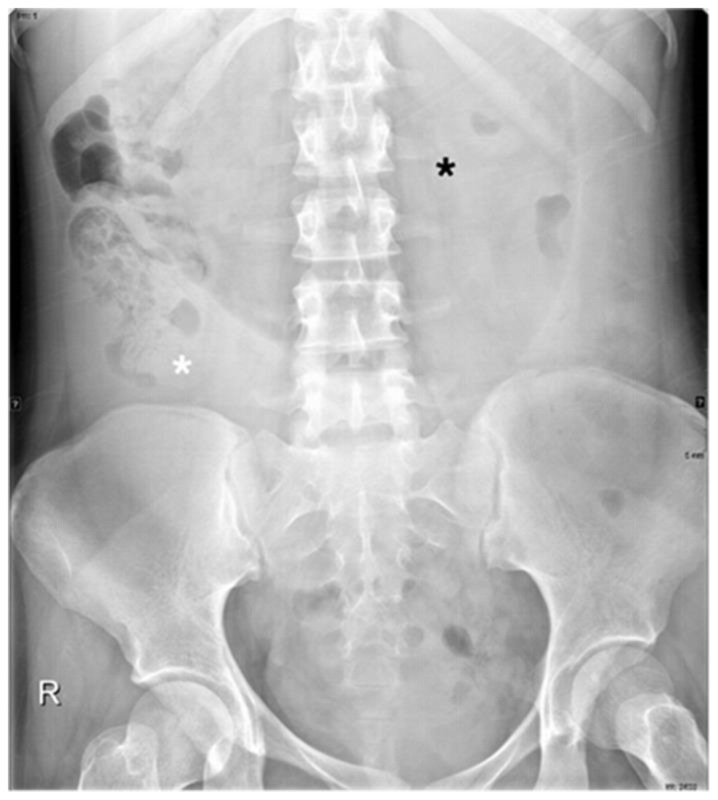
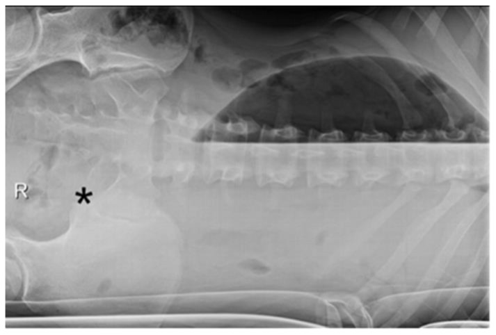
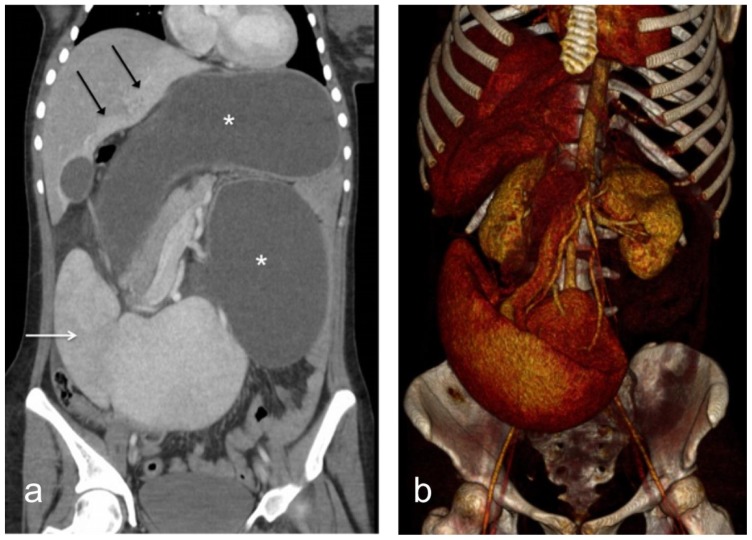
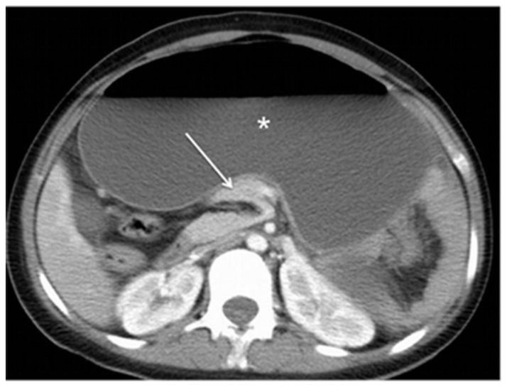
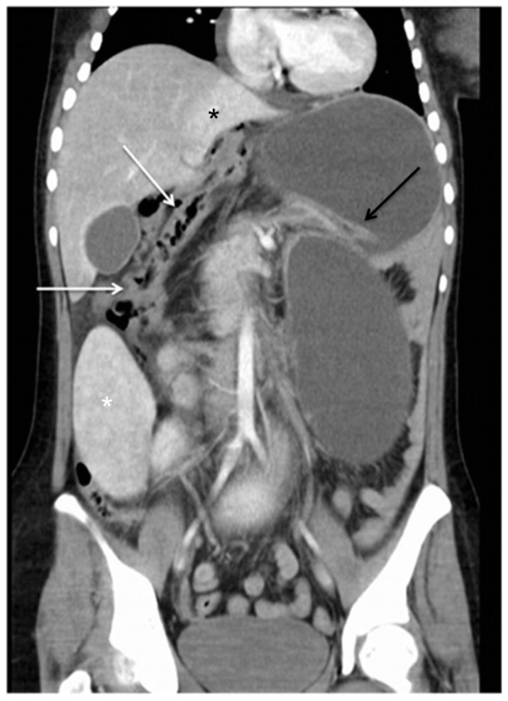
Wandering spleen is a rare condition, characterized by a mobile spleen that is attached only by an elongated vascular pedicle, allowing it to migrate to any part of the abdomen or pelvis. Mesenteroaxial gastric volvulus usually occurs in children and may be associated with wandering spleen. Both entities result from abnormal laxity or absence of the peritoneal attachments due to abnormal fusion of the peritoneal mesenteries. Pancreatic volvulus is a very rare anomaly, with only a few isolated case reports described in association with wandering spleen. Anomalous right sided descending and sigmoid colon is a very rare entity and its association with wandering spleen has not been previously reported. We report a case of wandering spleen associated with mesenteroaxial gastric volvulus, pancreatic volvulus and rightward shift of the splenic flexure of the colon and right sided descending and sigmoid colon in a young female.

Keywords: Colon; Computed tomography; Ectopic spleen; Gastric volvulus; Pancreatic volvulus; Wandering spleen.


Figures

Figure 1
Figure 1
A 22-year-old female with wandering spleen, gastric and pancreatic volvulus and right-sided descending colon. FINDINGS: Plain abdominal film shows a severe gastric distension (black asterisk), an empty splenic fossa and a mass in the right iliac fossa (white asterisk). TECHNIQUE: Supine abdominal plain film.
Figure 2
Figure 2
A 22-year-old female with wandering spleen, gastric and pancreatic volvulus and right-sided descending colon. FINDINGS: Plain abdominal film demonstrates a moveable mass in the left iliac fossa (black asterisk). TECHNIQUE: Left lateral decubitus abdominal film.
Figure 3
Figure 3
A 22-year-old female with wandering spleen, gastric and pancreatic volvulus and right-sided descending colon. FINDINGS: Coronal contrast-enhanced CT in portal venous phase (a) demonstrates a large spleen in the right iliac fossa with a perfusion defect in the upper pole (white arrow), a hyper-enhanced area of increased blood flow in the liver (black arrows) and a gastric and pancreatic volvulus (white asterisks). (b) 3D volume rendering image showing the spleen displaced into the right iliac fossa, the torsion of the splenic vessels and the pancreatic volvulus. TECHNIQUE: Axial contrast-enhanced CT. LightSpeed GE Medical Systems, kVp 120, mA 600, 5 mm slice thickness, Pitch 1.375:1, 100 cc of Ultravist 300 IV, in portovenous phase.
Figure 4
Figure 4
A 22-year-old female with wandering spleen, gastric and pancreatic volvulus and right-sided descending colon. FINDINGS: Axial post contrast CT scan in the portal venous phase showing the pancreatic volvulus (white arrow), markedly gastric distension and the absence of descending colon on the left flank. TECHNIQUE: Axial post contrast CT scan in the portal venous phase. LightSpeed GE Medical Systems, kVp 120, mA 600, 5 mm slice thickness, Pitch 1.375:1, 100 cc of Ultravist 300 IV, in portovenous phase.
Figure 5
Figure 5
A 22-year-old female with wandering spleen, gastric and pancreatic volvulus and right-sided descending colon. FINDINGS: Coronal contrast-enhanced CT in portal venous phase demonstrates a right-sided transverse and descending colon (white arrows). TECHNIQUE: Coronal post contrast CT scan in the portal venous phase LightSpeed GE Medical Systems, kVp 120, mA 600, 5 mm slice thickness, Pitch 1.375:1, 100 cc of Ultravist 300 IV, in portovenous phase.
Figure 6
Figure 6
A 22-year-old female with wandering spleen, gastric and pancreatic volvulus and right-sided descending colon. FINDINGS: Upper gastrointestinal endoscopy showing the distended stomach during the aspiration of liquid with fecaloid aspect (a), the torsioned pylorus (b) and the normal distal duodenum after devolvulation (c). TECHNIQUE: Flexible upper gastrointestinal endoscopy.
Figure 7
Figure 7
A 22-year-old female with wandering spleen, gastric and pancreatic volvulus and right-sided descending colon. FINDINGS: Abdominal plain film after endoscopic devolvulation shows the return of the spleen and transverse colon to a normal position (white asterisk) and resolution of the gastric volvulus. Note also the right-sided descending colon (white arrow). TECHNIQUE: Supine abdominal plain film.

References

    1. Raissaki M, Prassopoulos P, Daskalogiannaki M, Magkanas E, Gourtsoyiannis N. Acute abdomen due to torsion of wandering spleen: CT diagnosis. European radiology. 1998;8(8):1409–12. doi: 10.1007/s003300050562. - DOI - PubMed
    1. Ben Ely A, Zissin R, Copel L, Vasserman M, Hertz M, Gottlieb P, et al. The wandering spleen: CT findings and possible pitfalls in diagnosis. Clinical radiology. 2006;61(11):954–8. doi: 10.1016/j.crad.2006.06.007. - DOI - PubMed
    1. Liu HT, Lau KK. Wandering spleen: an unusual association with gastric volvulus. AJR American journal of roentgenology. 2007;188(4):W328–30. doi: 10.2214/AJR.05.0672. - DOI - PubMed
    1. Priyadarshi RN, Anand U, Kumar B, Prakash V. Torsion in wandering spleen: CT demonstration of whirl sign. Abdominal imaging. 2013;38(4):835–8. doi: 10.1007/s00261-012-9944-9. - DOI - PubMed
    1. Gorsi U, Bhatia A, Gupta R, Bharathi S, Khandelwal N. Pancreatic volvulus with wandering spleen and gastric volvulus: an unusual triad for acute abdomen in a surgical emergency. Saudi journal of gastroenterology : official journal of the Saudi Gastroenterology Association. 2014;20(3):195–8. doi: 10.4103/1319-3767.133026. - DOI - PMC - PubMed

Publication types

MeSH terms

LinkOut - more resources