

Accuracy of Platelet Counting by Optical and Impedance Methods in Patients with Thrombocytopaenia and Microcytosis
- PMID: 26629371
- PMCID: PMC4664089
- DOI: 10.18295/squmj.2015.15.04.004
Accuracy of Platelet Counting by Optical and Impedance Methods in Patients with Thrombocytopaenia and Microcytosis
Abstract
Objectives: Obtaining accurate platelet counts in microcytic blood samples is challenging, even with the most reliable automated haematology analysers. The CELL-DYN(™) Sapphire (Abbott Laboratories, Chicago, Illinois, USA) analyser uses both optical density and electronic impedance methods for platelet counting. This study aimed to evaluate the accuracy of optical density and electrical impedance methods in determining true platelet counts in thrombocytopaenic samples with microcytosis as defined by low mean corpuscular volume (MCV) of red blood cells. Additionally, the impact of microcytosis on platelet count accuracy was evaluated.
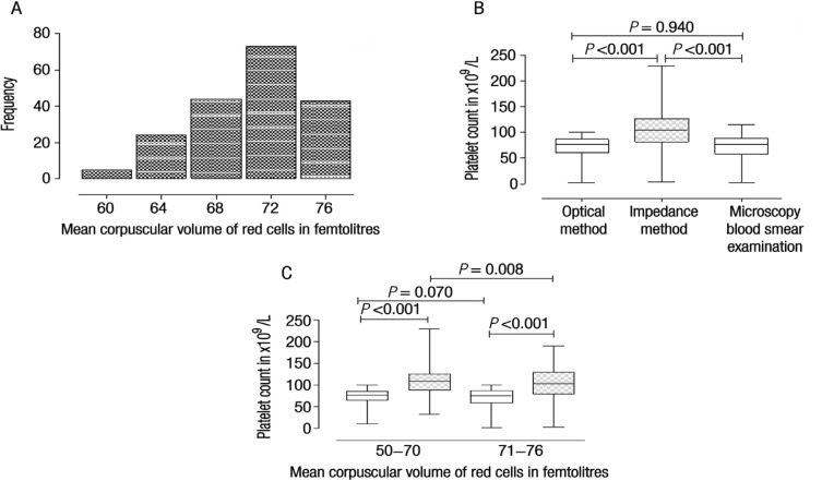
Methods: This study was carried out between February and December 2014 at the Haematology Laboratory of the Sultan Qaboos University Hospital in Muscat, Oman. Blood samples were collected and analysed from 189 patients with thrombocytopaenia and MCV values of <76 femtolitres. Platelet counts were tested using both optical and impedance methods. Stained peripheral blood films for each sample were then reviewed as a reference method to confirm platelet counts.
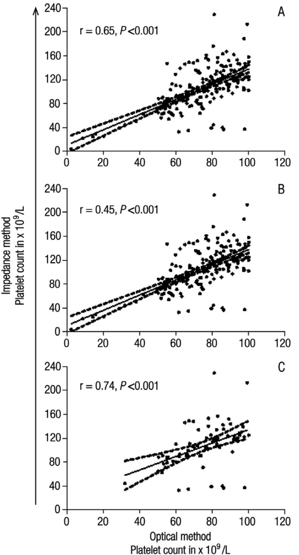
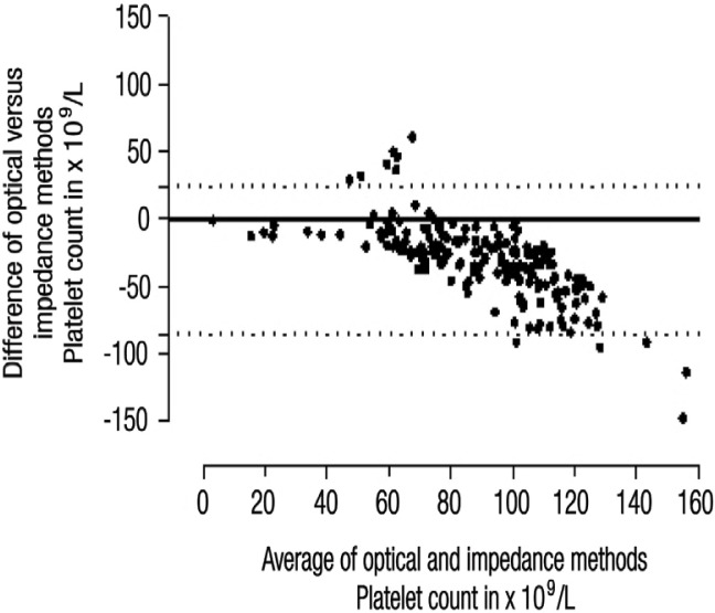
Results: The platelet counts estimated by the impedance method were on average 30% higher than those estimated by the optical method (P <0.001). The estimated intraclass correlation coefficient was 0.52 (95% confidence interval: 0.41-0.62), indicating moderate reliability between the methods. The degree of agreement between methods ranged from -85.5 to 24.3 with an estimated bias of -30, suggesting that these methods generate different platelet results.
Conclusion: The impedance method significantly overestimated platelet counts in microcytic and thrombocytopaenic blood samples. Further attention is therefore needed to improve the accuracy of platelet counts, particularly for patients with conditions associated with microcytosis.
Keywords: Anemia; Electrical Impedance; Mean Corpuscular Volume; Optical Devices; Platelet Counts; Thrombocytopenia.
Figures
References
-
- De la Salle BJ, McTaggart PN, Briggs C, Harrison P, Doré CJ, Longair I, et al. The accuracy of platelet counting in thrombocytopenic blood samples distributed by the UK National External Quality Assessment Scheme for General Haematology. Am J Clin Pathol. 2012;137:65–74. doi: 10.1309/AJCP86JMBFUCFCXA. - DOI - PubMed
-
- Cid J, Nascimento JD, Vicent A, Aguinaco R, Escoda L, Ugarriza A, et al. Evaluation of low platelet counts by optical, impedance, and CD61-immunoplatelet methods: Estimation of possible inappropriate platelet transfusion. Transfusion. 2010;50:795–800. doi: 10.1111/j.1537-2995.2009.02504.x. - DOI - PubMed
LinkOut - more resources
Full Text Sources
Other Literature Sources