. 2015 Aug 6;4(11):834-45.
doi: 10.1016/j.molmet.2015.07.009. eCollection 2015 Nov.

Human skeletal myotubes display a cell-autonomous circadian clock implicated in basal myokine secretion

Affiliations

Human skeletal myotubes display a cell-autonomous circadian clock implicated in basal myokine secretion

Laurent Perrin et al. Mol Metab. .

Abstract

Objective: Circadian clocks are functional in all light-sensitive organisms, allowing an adaptation to the external world in anticipation of daily environmental changes. In view of the potential role of the skeletal muscle clock in the regulation of glucose metabolism, we aimed to characterize circadian rhythms in primary human skeletal myotubes and investigate their roles in myokine secretion.

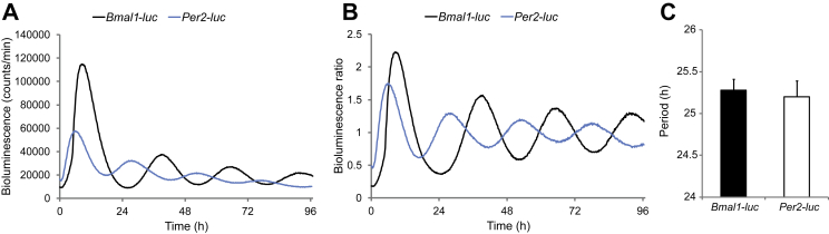
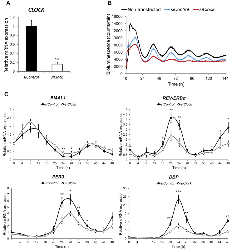
Methods: We established a system for long-term bioluminescence recording in differentiated human myotubes, employing lentivector gene delivery of the Bmal1-luciferase and Per2-luciferase core clock reporters. Furthermore, we disrupted the circadian clock in skeletal muscle cells by transfecting siRNA targeting CLOCK. Next, we assessed the basal secretion of a large panel of myokines in a circadian manner in the presence or absence of a functional clock.

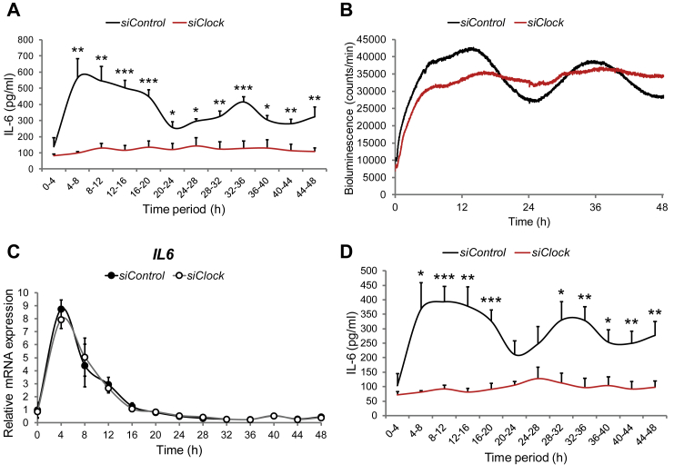
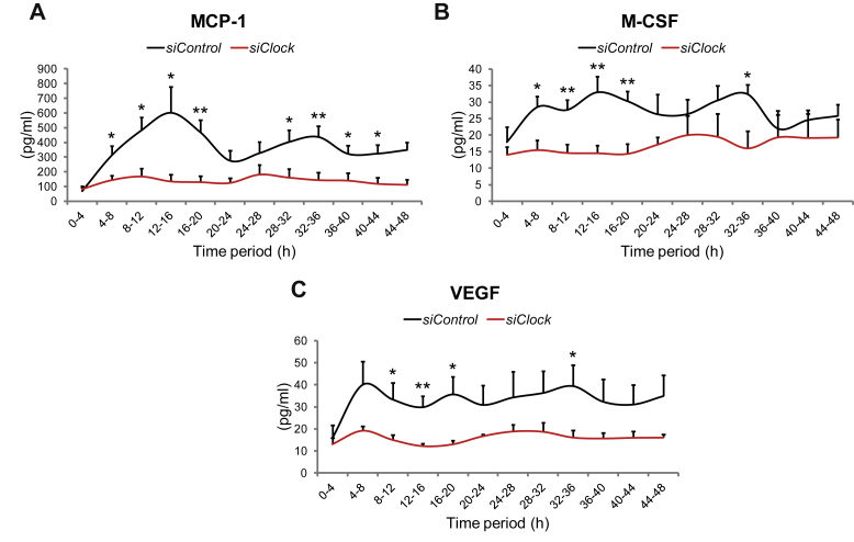
Results: Bioluminescence reporter assays revealed that human skeletal myotubes, synchronized in vitro, exhibit a self-sustained circadian rhythm, which was further confirmed by endogenous core clock transcript expression. Moreover, we demonstrate that the basal secretion of IL-6, IL-8 and MCP-1 by synchronized skeletal myotubes has a circadian profile. Importantly, the secretion of IL-6 and several additional myokines was strongly downregulated upon siClock-mediated clock disruption.

Conclusions: Our study provides for the first time evidence that primary human skeletal myotubes possess a high-amplitude cell-autonomous circadian clock, which could be attenuated. Furthermore, this oscillator plays an important role in the regulation of basal myokine secretion by skeletal myotubes.

Keywords: Circadian bioluminescence recording; Circadian clock; Human skeletal myotube; Interleukin-6; Myokine.


Figures

Figure 1
Figure 1
High-amplitude cell autonomous oscillators are functional in differentiated human primary myotubes. Human primary myoblasts were transduced with lentiviral particles expressing Bmal1-luc (black line) or Per2-luc (blue line). Cells were differentiated into myotubes, synchronized with forskolin, and transferred to the Actimetrics LumiCycle for bioluminescence recording. Raw (A) and detrended (B) oscillation profiles are representative of 19 and 14 independent experiments, respectively (one donor per experiment). (C) The period length of Bmal1-luc or Per2-luc was on average 25.29 ± 0.13 h, (n = 19) or 25.20 ± 0.19 h, (n = 14), respectively. Data represent the mean ± SEM.
Figure 2
Figure 2
Silencing of CLOCK expression attenuates circadian oscillations in human skeletal myotubes. (A) CLOCK mRNA was measured in human myotubes transfected with siControl or siClock by RT-qPCR and normalized to the mean of 9S and HPRT. CLOCK expression was reduced by 83.5 ± 3.4% (mean ± SEM, n = 8; ***p < 0.001) in siClock-transfected cells. (B) Amplitude of the Bmal1-luc reporter is strongly reduced in siClock-transfected myotubes. Representative Bmal1-luc oscillation profiles are shown for non-transfected (black line), siControl (blue line), and siClock (red line) transfected myotubes. Bmal1-luc oscillation profiles were recorded in duplicates (3 experiments, one donor per experiment). (C) RT-qPCR was performed for BMAL1, REV-ERBα, PER3 and DBP on RNA samples extracted from forskolin-synchronized human myotubes, transfected with siClock (open circles) or siControl (closed circles). Samples were collected every 4 h and normalized to the mean of 9S/HPRT. Profiles (mean ± SEM) are representative of 3 experiments (2 donors for time points 0 h–48 h and 3 donors for time points 12 h–36 h) with duplicates per time point.
Figure 3
Figure 3
Basal IL-6 secretion by human skeletal myotubes is strongly inhibited in the absence of a functional circadian clock. Myoblasts were transduced with the Bmal1-luc lentivector, differentiated into myotubes and transfected with either siControl or siClock siRNA. 24 h following transfection, myotubes were synchronized with forskolin and subjected to continuous perifusion with parallel bioluminescence recording. (A) Basal IL-6 secretion profile (mean + SEM) in the presence or absence of a functional clock. The perifusion outflow medium was collected continuously in an automated manner in 4 h intervals until 48 h (0–4 corresponds to the accumulation of IL-6 between 0 h and 4 h). IL-6 levels in the perifusion outflow medium were assessed by ELISA. The results represent basal IL-6 levels normalized to the total DNA content. 2 technical duplicates from 3 independent experiments (3 non-obese donors, see Table 3) were analyzed for each time point. (B) Bmal1-luc bioluminescence profiles of siControl-transfected myotubes (black line) and siClock-transfected myotubes (red line), representative of 3 experiments, with one donor cell line used per experiment. (C) RT-qPCR was performed for IL6 on RNA samples extracted from forskolin-synchronized human myotubes, transfected with siClock (open circles) or siControl (closed circles). Samples were collected every 4 h and normalized to the mean of 9S/HPRT. Profiles (mean ± SEM) are representative of 3 experiments (2 donors for time points 0 h–48 h and 3 donors for time points 12 h–36 h) with duplicates per time point. (D) Basal IL-6 secretion profiles in the presence or absence of a functional clock obtained from concentrated perifusion samples, assessed by multiplex analysis. Data shown as mean + SEM of 2 technical duplicates from 3 biological samples for each time point, normalized to the total DNA content.
Figure 4
Figure 4
Basal myokine secretion by human skeletal myotubes is affected by circadian clock disruption. Myoblasts were transduced with the Bmal1-luc lentivector, differentiated into myotubes, transfected with either siControl or siClock siRNA, and subjected to continuous perifusion with parallel bioluminescence recording. Concentrated perifusion samples were assessed by multiplex analysis. 2 technical duplicates from 3 biological samples were analyzed for each time point, and normalized to the total DNA content. Basal secretion profiles (mean + SEM) in the presence or absence of a functional clock are shown for (A) MCP-1, (B) M-CSF, and (C) VEGF.

References

    1. Albrecht U. Timing to perfection: the biology of central and peripheral circadian clocks. Neuron. 2012;74:246–260. - PubMed
    1. Bass J. Circadian topology of metabolism. Nature. 2012;491:348–356. - PubMed
    1. Dibner C., Schibler U. Circadian timing of metabolism in animal models and humans. Journal of Internal Medicine. 2015 May;277(5):513–527. - PubMed
    1. Marcheva B., Ramsey K.M., Peek C.B., Affinati A., Maury E., Bass J. Circadian clocks and metabolism. Handbook of Experimental Pharmacology. 2013:127–155. - PMC - PubMed
    1. Damiola F., Le Minh N., Preitner N., Kornmann B., Fleury-Olela F., Schibler U. Restricted feeding uncouples circadian oscillators in peripheral tissues from the central pacemaker in the suprachiasmatic nucleus. Genes & Development. 2000;14:2950–2961. - PMC - PubMed

LinkOut - more resources