. 2015 Aug 4;2(10):1420-9.
doi: 10.1016/j.ebiom.2015.08.002. eCollection 2015 Oct.

Compensation in Preclinical Huntington's Disease: Evidence From the Track-On HD Study

Collaborators, Affiliations

Compensation in Preclinical Huntington's Disease: Evidence From the Track-On HD Study

Stefan Klöppel et al. EBioMedicine. .

Abstract

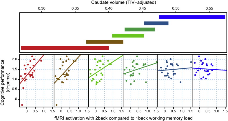
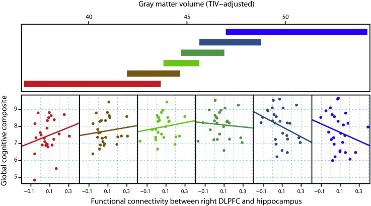
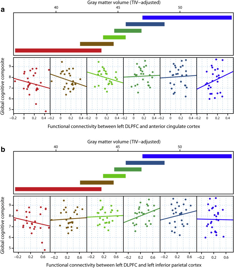
Background: Cognitive and motor task performance in premanifest Huntington's disease (HD) gene-carriers is often within normal ranges prior to clinical diagnosis, despite loss of brain volume in regions involved in these tasks. This indicates ongoing compensation, with the brain maintaining function in the presence of neuronal loss. However, thus far, compensatory processes in HD have not been modeled explicitly. Using a new model, which incorporates individual variability related to structural change and behavior, we sought to identify functional correlates of compensation in premanifest-HD gene-carriers.

Methods: We investigated the modulatory effects of regional brain atrophy, indexed by structural measures of disease load, on the relationship between performance and brain activity (or connectivity) using task-based and resting-state functional MRI.

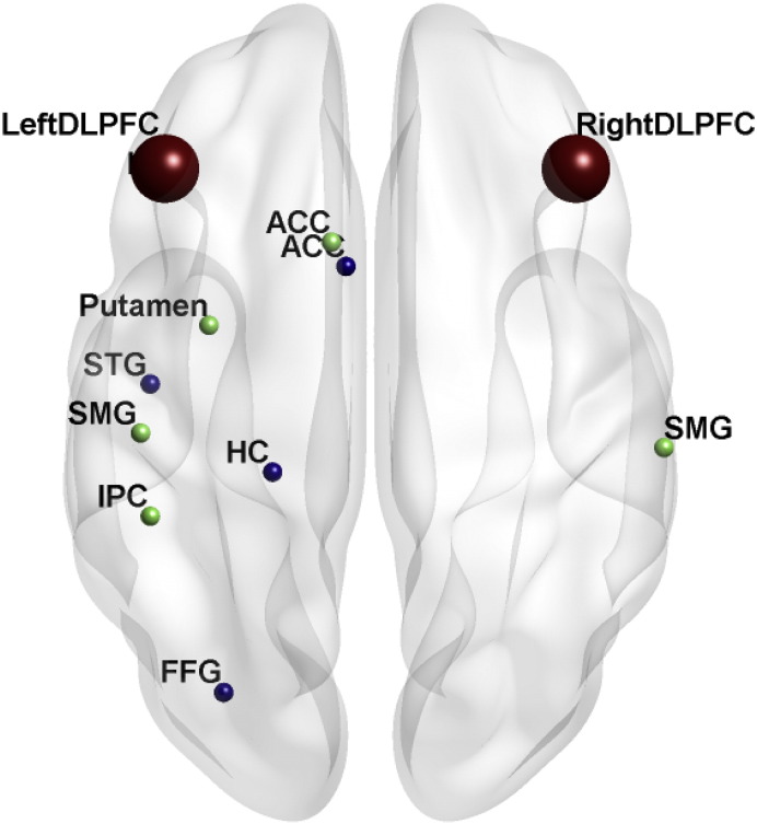
Findings: Consistent with compensation, as atrophy increased performance-related activity increased in the right parietal cortex during a working memory task. Similarly, increased functional coupling between the right dorsolateral prefrontal cortex and a left hemisphere network in the resting-state predicted better cognitive performance as atrophy increased. Such patterns were not detectable for the left hemisphere or for motor tasks.

Interpretation: Our findings provide evidence for active compensatory processes in premanifest-HD for cognitive demands and suggest a higher vulnerability of the left hemisphere to the effects of regional atrophy.

Keywords: Cognitive; Huntington's disease; MRI; Motor; Neural compensation; Preclinical.


Figures

Fig. 1
Fig. 1
Example conditioning plot with simulated data. The upper panel depicts the overlapping ranges of structural disease load as measured by brain volume (the slabs) that determine the subsample for each scatterplot panel below. Observed points are plotted in the lower panel scatterplots for each range of brain volume, and a linear regression line is fit separately in each panel to aid interpretation. The structural disease load (brain volume) range determines what subsample is selected from the data set for the scatterplot of task performance as a function of fMRI signal, with the color coding showing the correspondence. For example, the extreme left scatterplot (red) includes the smallest brain volume (highest disease load) range from the data set (lower left red slab). The extreme right scatterplot (blue) includes the largest volume (lowest disease load) range from the data set (upper right blue slab). Of note, regression lines and the separation in different slabs have illustrative purposes only and are not the basis of the underlying statistic.
Fig. 2
Fig. 2
Task fMRI paradigms. Example trials for a) the verbal working memory n-back task and b) sequential finger tapping. Please see Materials and Methods for further details.
Fig. 3
Fig. 3
Dynamic causal models employed for resting state fMRI. a) Motor network and b) cognitive network. Abbreviations: ACC: anterior cingulate cortex; DLPFC: dorsolateral prefrontal cortex; PMC: premotor cortex; PPC: posterior parietal cortex; and SMA: supplementary motor area.
Fig. 4
Fig. 4
D-Prime performance as a function of fMRI task activation within the parietal cortex, conditional on caudate volume as a measure of structural disease load. For each plot, the upper panel depicts the overlapping ranges of caudate volume that determine which subsample is selected from the data set that is used to construct each scatterplot. A linear regression line was fit for each scatterplot to aid interpretation.
Fig. 5
Fig. 5
Overview of functional connectivity analyses for the cognitive network. Regions that significantly correlated (p < 0.05 FWE-corrected) with seed regions in the right (blue) or left DLPFC (green) and which also, as part of the compensation model, significantly predicted global cognitive performance as structural disease load increased. Abbreviations: ACC: anterior cingulate cortex; DLPFC: dorsolateral prefrontal cortex; FFG: fusiform gyrus; HC: hippocampus; IPC: inferior parietal cortex; and SMG: supramarginal gyrus. STG: superior temporal gyrus.
Fig. 6
Fig. 6
Global cognitive performance as a function of rsfMRI functional connectivity between right DLPFC and left hippocampus, conditional on gray matter volume as a measure of structural disease load. For each plot, the upper panel depicts the overlapping ranges of gray matter volume that determine which subsample is selected from the data set that is used to construct each scatterplot. A linear regression line was fit for each scatterplot to aid interpretation.
Fig. 7
Fig. 7
Global cognitive performance as a function of rsfMRI functional connectivity between left DLPFC and a) anterior cingulate cortex, or b) left inferior parietal cortex, conditional on gray matter volume as a measure of structural disease load. For each plot, the upper panel depicts the overlapping ranges of gray matter volume that determine which subsample is selected from the data set that is used to construct each scatterplot. A linear regression line was fit for each scatterplot to aid interpretation.

Comment in

References

    1. Ashburner J. A fast diffeomorphic image registration algorithm. NeuroImage. 2007;38:95–113. - PubMed
    1. Barulli D., Stern Y. Efficiency, capacity, compensation, maintenance, plasticity: emerging concepts in cognitive reserve. Trends Cogn. Sci. 2013;17:502–509. - PMC - PubMed
    1. Beckmann C.F., DeLuca M., Devlin J.T., Smith S.M. Investigations into resting-state connectivity using independent component analysis. Philos. Trans. R. Soc. Lond. B Biol. Sci. 2005;360:1001–1013. - PMC - PubMed
    1. Button K.S., Ioannidis J.P.A., Mokrysz C. Power failure: why small sample size undermines the reliability of neuroscience. Nat. Rev. Neurosci. 2013;14:365–376. - PubMed
    1. Cabeza R.E., Dennis N.A. Frontal lobes and aging: deterioration and compensation. In: Stuss D.T., Knight R.T., editors. Principles of Frontal Lobe Function. 2nd edn. Oxford University Press; New York: 2013. pp. 628–652.

Publication types