. 2015 Jul 31;2(10):1536-48.
doi: 10.1016/j.ebiom.2015.07.040. eCollection 2015 Oct.

Pathophysiologic Response to Burns in the Elderly

Affiliations

Pathophysiologic Response to Burns in the Elderly

Marc G Jeschke et al. EBioMedicine. .

Abstract

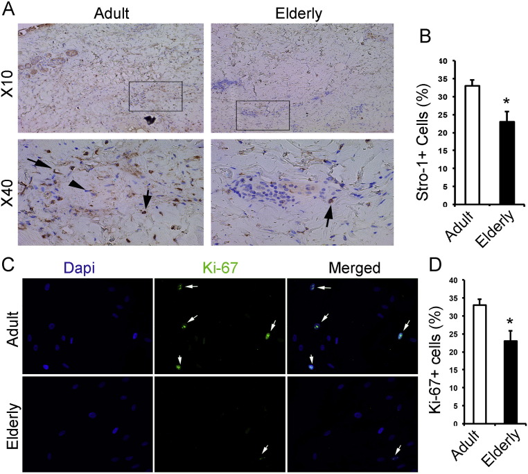
Over the last decades advancements have improved survival and outcomes of severely burned patients except one population, elderly. The Lethal Dose 50 (LD50) burn size in elderly has remained the same over the past three decades, and so has morbidity and mortality, despite the increased demand for elderly burn care. The objective of this study is to gain insights on why elderly burn patients have had such a poor outcome when compared to adult burn patients. The significance of this project is that to this date, burn care providers recognize the extreme poor outcome of elderly, but the reason remains unclear. In this prospective translational trial, we have determined clinical, metabolic, inflammatory, immune, and skin healing aspects. We found that elderly have a profound increased mortality, more premorbid conditions, and stay at the hospital for longer, p < 0.05. Interestingly, we could not find a higher incidence of infection or sepsis in elderly, p > 0.05, but a significant increased incidence of multi organ failure, p < 0.05. These clinical outcomes were associated with a delayed hypermetabolic response, increased hyperglycemic and hyperlipidemic responses, inversed inflammatory response, immune-compromisation and substantial delay in wound healing predominantly due to alteration in characteristics of progenitor cells, p < 0.05. In summary, elderly have substantially different responses to burns when compared to adults associated with increased morbidity and mortality. This study indicates that these responses are complex and not linear, requiring a multi-modal approach to improve the outcome of severely burned elderly.

Keywords: Burn; Elderly; Hypermetabolism; Inflammasome; Inflammation; Morbidity; Mortality; Pathophysiologic response; Skin healing; Stem cell.


Figures

Supplemental Fig. 1
Supplemental Fig. 1
Oral Glucose Tolerance Test (OGTT) glucose, insulin, and c-peptide. Oral Glucose Tolerance test were performed on patients and glucose (A), insulin (C) and c-Peptide (E) quantified. The respective AUC for these markers are shown in (B), (D) and (F). n for adults = 55 and for elderly = 29* Significant difference between adults vs. elderly, p < 0.05.
Supplemental Fig. 2
Supplemental Fig. 2
Lipid profile of adult and elderly burned patients. Serum free fatty acid species were analyzed by GCMS and a heat map was created to illustrate their concentration sin the respective groups of patients (A). The sum of the FFAs and the percentage of fatty acids subfamilies are shown in B and C respectively. n for control = 5, for adults = 10 and for elderly = 10. Data expressed as absolute values or as mean ± SEM. * Significant difference between control vs. adult, p < 0.05. §Significant difference between control vs. elderly, p < 0.05. φ Significant difference between adults vs. elderly, p < 0.05. Using double symbols means p < 0.001.
Supplemental Fig. 3
Supplemental Fig. 3
Metabolic profile over time. Adult and elderly have increased c-Peptide and GLP-1 levels, as well as decreased (Pancreatic Peptide) PP, (Peptide YY) PYY, Leptin, GIP and Ghrelin levels (A–J). Data is represented as mean ± SEM. Significant difference between adults vs. elderly, *p < 0.05 and **p < 0.01.
Supplemental Fig. 4
Supplemental Fig. 4
Inflammasome in WAT. Elderly patients have less macrophages (A) and Interleukin-1 beta (IL-1β +) cells in the CD14 + macrophage fraction (B–C), p > 0.05. In the fraction of CD14 + macrophage that are double-positive for IL-1β and FLICA (caspase-1), both adults and elderly were significantly higher than healthy controls (D). Both length of stay and Baux score correlated with the percentage of IL-1β +, CD14 + cells (E–F). n for adult = 18 and for elderly = 11.
Supplemental Fig. 5
Supplemental Fig. 5
Altered expression of Oct-4 transcription factor in the mesenchymal cells isolated from skin of elderlies in compare with young adults. (A–B) Representative fluorescence images of cells from burned human skin of elderly patients show reduced number of cells positive for Oct4 (green). DAPI (4,6-diamidino-2-phenylindole) was used to identify the nuclei (blue). Arrows indicate cells positive for nuclear Oct4. (n = 3 for each group).
Fig. 1
Fig. 1
Kaplan Meier survival curves. (A) Survival curve for adults vs. elderly from 1995 to 2015 for all patients and all burn sizes. (B) Survival curve for burn patients with burns over 20% total body surface area (TBSA) adults vs. elderly from 1995 to April 2015. (C) Survival curve for adults vs. elderly from 2006 to April 2015 for all patients all burn sizes. (D) Survival curve for burn patients with burns over 20% TBSA adults vs. elderly from 2006 to April 2015. * Significant difference between adults vs. elderly p < 0.05.
Fig. 2
Fig. 2
Metabolic measurements. (A) Resting energy expenditure expressed as percent predicted was significantly increased in elderly patients > 4 weeks after burn when compared to adults. * Significant difference between adults vs. elderly, p < 0.05. (B) 6 AM average glucose levels. Elderly have a significantly greater area under the curve (AUC) at 6 AM glucose levels compared to adults. (C) Daily average glucose levels. Elderly have a significantly greater AUC when compared to adults. (D) Daily max glucose levels were also significantly higher when compared to adults. (E) No differences could be detected between elderly and adults in daily min glucose levels. (F) Significantly elderly required more insulin than adults.
Fig. 2
Fig. 2
Metabolic measurements. (A) Resting energy expenditure expressed as percent predicted was significantly increased in elderly patients > 4 weeks after burn when compared to adults. * Significant difference between adults vs. elderly, p < 0.05. (B) 6 AM average glucose levels. Elderly have a significantly greater area under the curve (AUC) at 6 AM glucose levels compared to adults. (C) Daily average glucose levels. Elderly have a significantly greater AUC when compared to adults. (D) Daily max glucose levels were also significantly higher when compared to adults. (E) No differences could be detected between elderly and adults in daily min glucose levels. (F) Significantly elderly required more insulin than adults.
Fig. 3
Fig. 3
Metabolic bio-molecular markers. (A–D) Adipose ER stress markers. Adults and elderly both increased adipose ER stress after burn, elderly burn patients had a significantly decreased ER stress response in the adipose tissue when compared with adult burn patients. (E–F) pJNK-JNK and NLRP3 and found that both adults and elderly have similar expression patterns with significantly increased levels at early and late time points with no significant difference between adults and elderly. (G–I) MPO in adipose tissue. Elderly have significantly lower MPO expression in adipose at 0–3 days and 14–20 days after burn when compared with adults indicating less inflammatory infiltration of the adipose tissue. n = 5 to 8 per group. Data is represented as mean ± SEM. * Significant difference between adults vs. elderly, p < 0.05.
Fig. 4
Fig. 4
Inflammatory and immune profile in adult and elderly burned patients. (A–F) Adults show a decrease in inflammatory mediators over time, while elderly demonstrate the opposite picture; IL-6, TNF, IL-15, MCP-1 and GM–CSF all increased over the time course after burn indicating a hypo-inflammation followed by hyper-inflammation. (G–J) Elderly had significantly reduced numbers of CD14 +/HLA-DR + monocytes (G). Comparing septicemia patients exclusively, the impaired phenotype previously observed was upheld (H). Despite a relatively steady impairment in CD14 +/HLA-DR + monocytes expression in adults, elderly patients had a notable surge in at 30 + days post-injury. Lastly, severely burned (> 30% TBSA) elderly patients showed a correlation between HLA-DR + expression and length of stay (I–J). n for analysis of inflammatory cytokine: adult = 94 and elderly = 36. n for HLA-DR flow cytometry: healthy = 11, adult = 37 and elderly 15. Data is represented as mean ± SEM. * Significant difference between adults vs. elderly, p < 0.05.
Fig. 5
Fig. 5
Deficient characteristics of skin progenitor cells in elderly patients. (A–B) Representative immunohistochemistry staining is showing that skin tissue elderly patients has less Stro-1 + cells. (C–D) Individual signals of 4,6-diamidino-2-phenylindole (DAPI) and anti-Ki-67 (green) from isolated skin cells of young adults and elderly burned patients are shown on alongside a merged view of DAPI. (E–F) Representative photomicrographs of a scratch assay performed with young adult skin mesenchymal cells and elderly's skin mesenchymal cells after 48 h. Migration was quantified by counting the number of cells migrating into the scratch zone. (G) Western blot showing lower activation (quantified in I) of Wnt/β-catenin signaling in the burned skin of elderly in compare with young adult. (H) Western blot showing lower activation (quantified in J) of TGF-β/Smad2 pathway in the burned skin of elderly in compare with young adult. Bar graphs show the means and standard deviation (n = 3–5). Three representative lysates of 3 subjects have been shown here. Data is represented as mean ± SEM. * Significant difference between adults vs. elderly, p < 0.05 and ** indicates p < 0.01.
Fig. 5
Fig. 5
Deficient characteristics of skin progenitor cells in elderly patients. (A–B) Representative immunohistochemistry staining is showing that skin tissue elderly patients has less Stro-1 + cells. (C–D) Individual signals of 4,6-diamidino-2-phenylindole (DAPI) and anti-Ki-67 (green) from isolated skin cells of young adults and elderly burned patients are shown on alongside a merged view of DAPI. (E–F) Representative photomicrographs of a scratch assay performed with young adult skin mesenchymal cells and elderly's skin mesenchymal cells after 48 h. Migration was quantified by counting the number of cells migrating into the scratch zone. (G) Western blot showing lower activation (quantified in I) of Wnt/β-catenin signaling in the burned skin of elderly in compare with young adult. (H) Western blot showing lower activation (quantified in J) of TGF-β/Smad2 pathway in the burned skin of elderly in compare with young adult. Bar graphs show the means and standard deviation (n = 3–5). Three representative lysates of 3 subjects have been shown here. Data is represented as mean ± SEM. * Significant difference between adults vs. elderly, p < 0.05 and ** indicates p < 0.01.

Comment in

References

    1. (WHO) OWH . 2002. A Graphical Overview of the Global Burden of Injuries. The Injury Chart Book. (Geneva)
    1. Abdullahi A., Amini-Nik S., Jeschke M.G. Animal models in burn research. Cell. Mol. Life Sci. 2014;71:3241–3255. - PMC - PubMed
    1. Albornoz C.R., Villegas J., Sylvester M., Pena V., Bravo I. Burns are more aggressive in the elderly: proportion of deep burn area/total burn area might have a role in mortality. Burns. 2011;37:1058–1061. - PubMed
    1. Amini Nik S., Ebrahim R.P., Van Dam K., Cassiman J.J., Tejpar S. TGF-beta modulates beta-catenin stability and signaling in mesenchymal proliferations. Exp. Cell Res. 2007;313:2887–2895. - PubMed
    1. Amini-Nik S., Glancy D., Boimer C., Whetstone H., Keller C., Alman B.A. Pax7 expressing cells contribute to dermal wound repair, regulating scar size through a beta-catenin mediated process. Stem Cells (Dayton, Ohio) 2011;29:1371–1379. - PubMed

MeSH terms