. 2015 Dec 2;10(12):e0143809.
doi: 10.1371/journal.pone.0143809. eCollection 2015.

Evaluating Models of Cellulose Degradation by Fibrobacter succinogenes S85

Affiliations

Evaluating Models of Cellulose Degradation by Fibrobacter succinogenes S85

Meagan C Burnet et al. PLoS One. .

Abstract

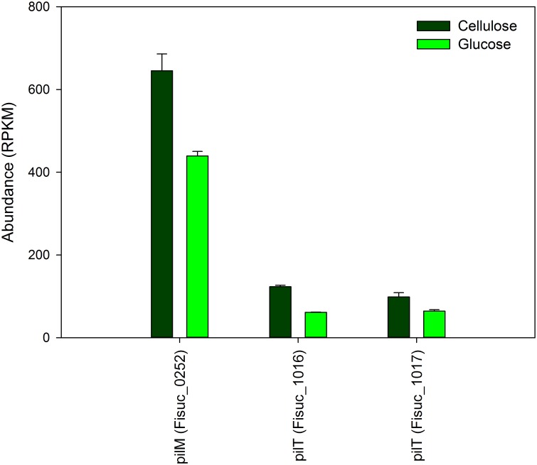
Fibrobacter succinogenes S85 is an anaerobic non-cellulosome utilizing cellulolytic bacterium originally isolated from the cow rumen microbial community. Efforts to elucidate its cellulolytic machinery have resulted in the proposal of numerous models which involve cell-surface attachment via a combination of cellulose-binding fibro-slime proteins and pili, the production of cellulolytic vesicles, and the entry of cellulose fibers into the periplasmic space. Here, we used a combination of RNA-sequencing, proteomics, and transmission electron microscopy (TEM) to further clarify the cellulolytic mechanism of F. succinogenes. Our RNA-sequence analysis shows that genes encoding type II and III secretion systems, fibro-slime proteins, and pili are differentially expressed on cellulose, relative to glucose. A subcellular fractionation of cells grown on cellulose revealed that carbohydrate active enzymes associated with cellulose deconstruction and fibro-slime proteins were greater in the extracellular medium, as compared to the periplasm and outer membrane fractions. TEMs of samples harvested at mid-exponential and stationary phases of growth on cellulose and glucose showed the presence of grooves in the cellulose between the bacterial cells and substrate, suggesting enzymes work extracellularly for cellulose degradation. Membrane vesicles were only observed in stationary phase cultures grown on cellulose. These results provide evidence that F. succinogenes attaches to cellulose fibers using fibro-slime and pili, produces cellulases, such as endoglucanases, that are secreted extracellularly using type II and III secretion systems, and degrades the cellulose into cellodextrins that are then imported back into the periplasm for further digestion by β-glucanases and other cellulases.


Conflict of interest statement

Competing Interests: The authors have declared that no competing interests exist.

Figures

Fig 1
Fig 1. Expression of fibro-slime genes in F. succinogenes S85 cellulose and glucose cultures.
The cellulose cultures showed an increased expression for 8 out of 10 fibro-slime proteins relative to the glucose cultures. RPKM = Reads Per Kilobase per Million mapped reads.
Fig 2
Fig 2. Expression of pilin transcripts in F. succinogenes S85 cellulose and glucose cultures.
There is a higher expression of pilin proteins in the cellulose cultures than the glucose cultures for 3 out of the 4 pilin proteins observed. RPKM = Reads Per Kilobase per Million mapped reads.
Fig 3
Fig 3. Difference in expression between cellulose and glucose cultures for transporters, secretion systems, effluxes, and exporters genes.
All type II and III secretion systems had higher expression in the cellulose cultures than the glucose cultures suggesting they may have a role in secreting the carbohydrate active enzymes into the extracellular medium. RPKM = Reads Per Kilobase per Million mapped reads.
Fig 4
Fig 4. (A) Average number of carbohydrate active enzymes (CAZymes) and (B) average abundance of CAZymes observed from a given fraction. (C) Percent of CAZymes to total proteins and (D) percent CAZymes abundance to total protein abundance.
The number and percentages of CAZymes and abundance of CAZymes was largest for the extracellular medium fraction. EM = extracellular medium, OM = outer membrane, PE = periplasm.
Fig 5
Fig 5. Comparison of the number of identified proteins and their overlap observed in the outer membrane, periplasm and extracellular medium fractions.
Fig 6
Fig 6. Average abundance of fibro-slime proteins identified from each fractionation.
The average abundance for all fibro-slime proteins was the highest in the extracellular medium.
Fig 7
Fig 7. TEM images of F. succinogenes S85 harvested during mid-exponential growth on cellulose (A, B), cellulose stationary growth (C, D), glucose mid-exponential growth (E) and glucose stationary growth (F).
Grooves in the cellulose were observed in the cellulose mid-exponential and stationary phases. Vesicles were present in only the cellulose stationary phase (arrows in D). Scale bars are 1 μm (A,C) and 0.5 μm (B-F). Vesicles are indicated by arrows. Grooves are indicated by curves.

References

    1. Bera-Maillet C, Ribot Y, Forano E. Fiber-degrading systems of different strains of the genus Fibrobacter. Appl Environ Microb. 2004;70(4):2172–9. 10.1128/Aem.70.4.2172-2179.2004 WOS:000220792200034. - DOI - PMC - PubMed
    1. Koike S, Yoshitani S, Kobayashi Y, Tanaka K. Phylogenetic analysis of fiber-associated rumen bacterial community and PCR detection of uncultured bacteria. Fems Microbiol Lett. 2003;229(1):23–30. 10.1016/S0378-1097(03)00760-2 WOS:000187362100004. - DOI - PubMed
    1. Suen G, Weimer PJ, Stevenson DM, Aylward FO, Boyum J, Deneke J, et al. The Complete Genome Sequence of Fibrobacter succinogenes S85 Reveals a Cellulolytic and Metabolic Specialist. Plos One. 2011;6(4). ARTN e18814 10.1371/journal.pone.0018814 WOS:000289671100023. - DOI - PMC - PubMed
    1. Groleau D, Forsberg CW. Cellulolytic Activity of the Rumen Bacterium Bacteroides-Succinogenes. Can J Microbiol. 1981;27(5):517–30. WOS:A1981LP27900007. - PubMed
    1. Forsberg CW, Beveridge TJ, Hellstrom A. Cellulase and Xylanase Release from Bacteroides-Succinogenes and Its Importance in the Rumen Environment. Appl Environ Microb. 1981;42(5):886–96. WOS:A1981MQ52800022. - PMC - PubMed

Publication types

LinkOut - more resources