. 2015 Dec 29;112(52):15862-7.
doi: 10.1073/pnas.1520099112. Epub 2015 Nov 16.

Augmentor α and β (FAM150) are ligands of the receptor tyrosine kinases ALK and LTK: Hierarchy and specificity of ligand-receptor interactions

Affiliations

Augmentor α and β (FAM150) are ligands of the receptor tyrosine kinases ALK and LTK: Hierarchy and specificity of ligand-receptor interactions

Andrey V Reshetnyak et al. Proc Natl Acad Sci U S A. .

Abstract

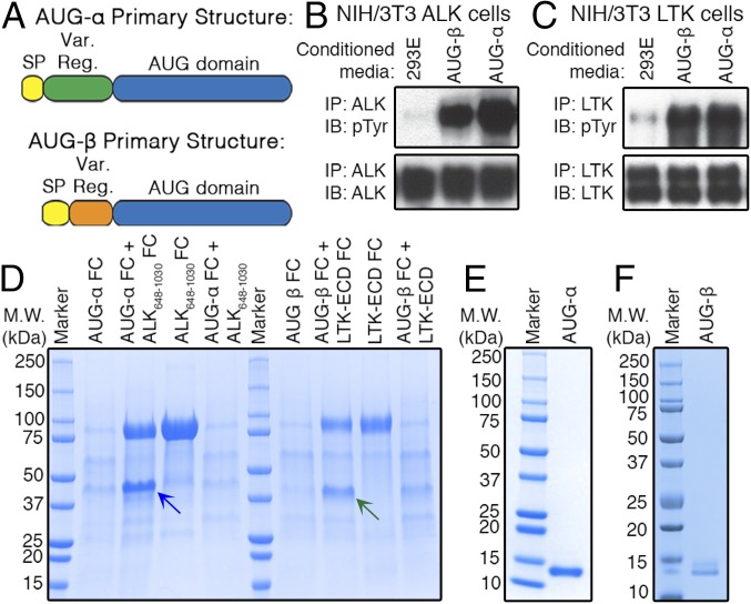
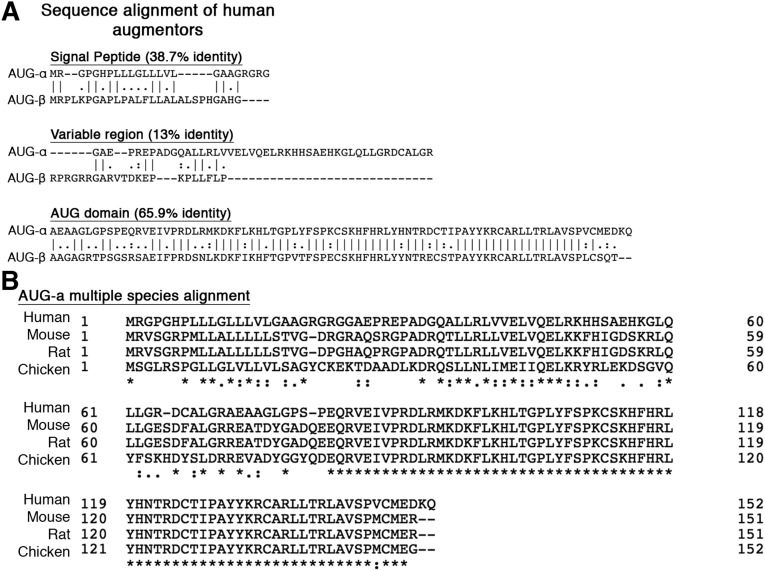
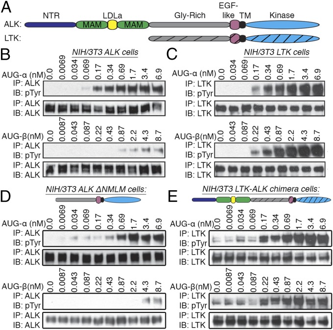
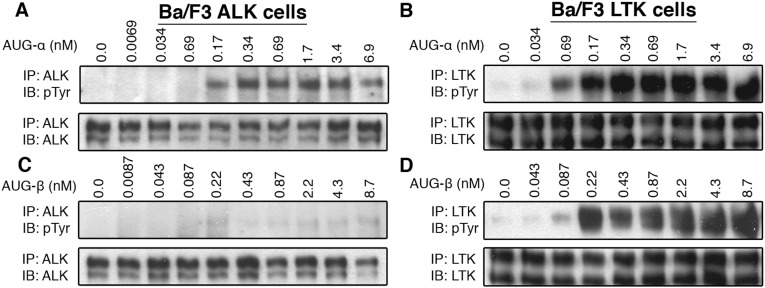
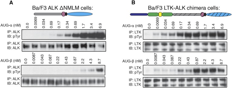
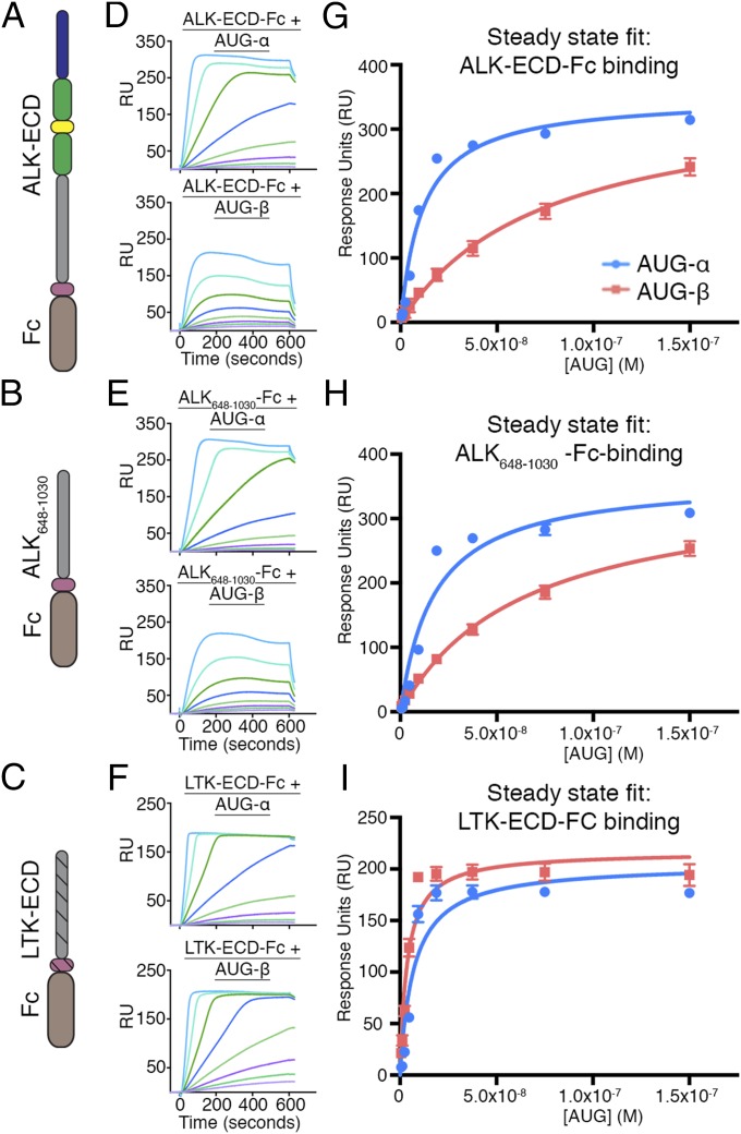
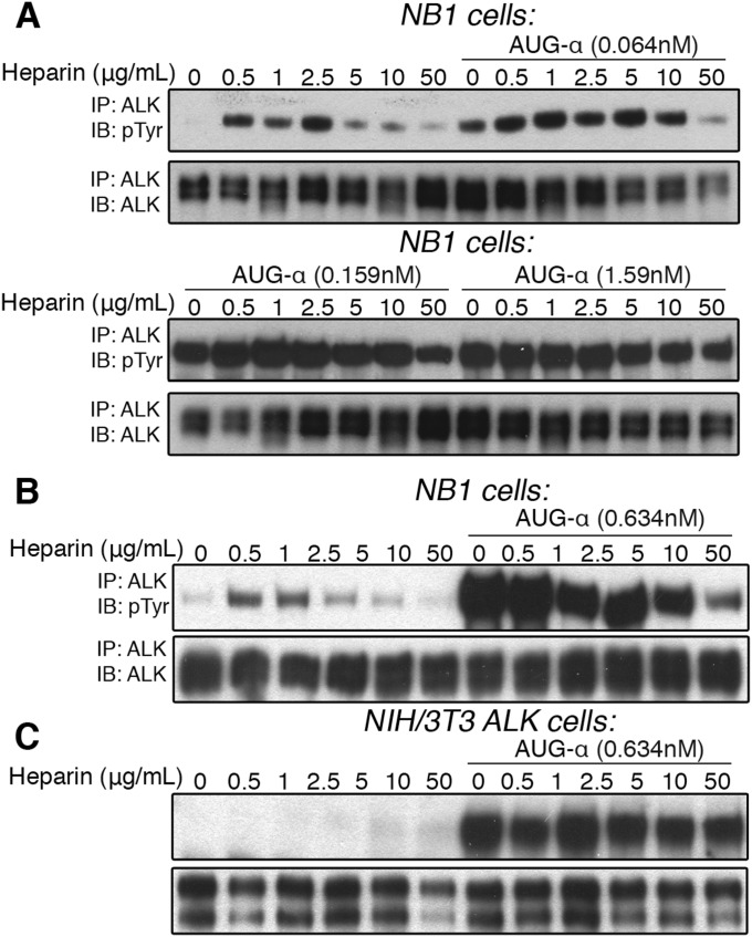
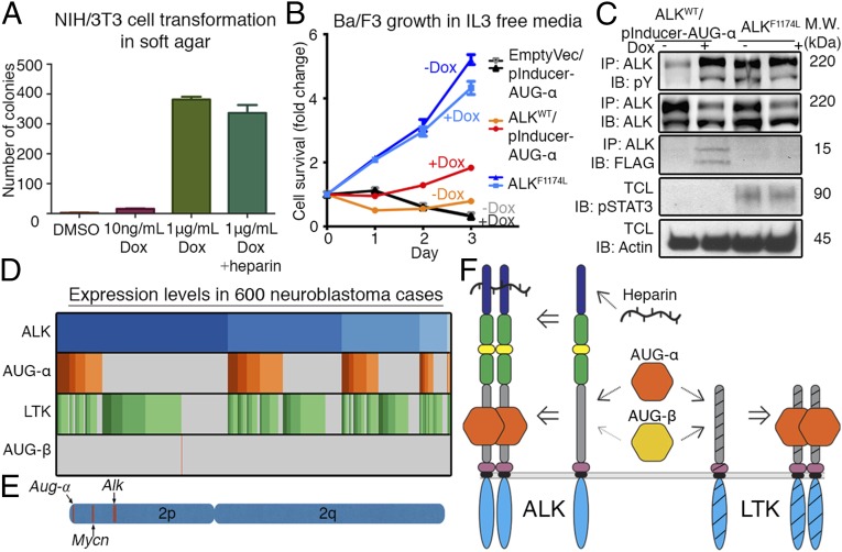
Receptor tyrosine kinases (RTKs) are a class of cell surface receptors that, upon ligand binding, stimulate a variety of critical cellular functions. The orphan receptor anaplastic lymphoma kinase (ALK) is one of very few RTKs that remain without a firmly established protein ligand. Here we present a novel cytokine, FAM150B, which we propose naming augmentor-α (AUG-α), as a ligand for ALK. AUG-α binds ALK with high affinity and activates ALK in cells with subnanomolar potency. Detailed binding experiments using cells expressing ALK or the related receptor leukocyte tyrosine kinase (LTK) demonstrate that AUG-α binds and robustly activates both ALK and LTK. We show that the previously established LTK ligand FAM150A (AUG-β) is specific for LTK and only weakly binds to ALK. Furthermore, expression of AUG-α stimulates transformation of NIH/3T3 cells expressing ALK, induces IL-3 independent growth of Ba/F3 cells expressing ALK, and is expressed in neuroblastoma, a cancer partly driven by ALK. These experiments reveal the hierarchy and specificity of two cytokines as ligands for ALK and LTK and set the stage for elucidating their roles in development and disease states.

Keywords: cancer; cell signaling; phosphorylation; protein kinases; surface receptors.


Conflict of interest statement

The authors declare no conflict of interest.

Figures

Fig. 1.
Fig. 1.
AUG-α and AUG-β are ligands for ALK and LTK. (A) Schematic representation of AUG-α and AUG-β. Signal peptide (SP), yellow; variable region (Var. Reg.), green (AUG-α) and orange (AUG-β); conserved AUG domain, blue. (B and C) NIH/3T3 cells stably expressing ALK (B) or LTK (C) were stimulated with conditioned medium from 293-E cells (negative control), or 293-E cells expressing AUG-α or AUG-β. Lysates of 3T3 cells were subjected to immunoprecipitation (IP) using anti-ALK (B) or anti-LTK (C) antibodies followed by SDS/PAGE and immunoblotting (IB) with anti-pTyr (IB: pTyr) or anti-ALK (IB: ALK) or anti-LTK (IB: LTK) antibodies (as indicated). (D) Expression of human augmentor ligands. AUG-α, fused to Fc fragment at the C terminus (AUG-α-Fc), was expressed alone or coexpressed with ALK648–1,030 or ALK648–1,030-Fc fusion protein. Similarly, fusion of AUG-β-Fc was expressed alone or coexpressed with LTK-ECD-Fc or with LTK-ECD. Coexpression of ALK648–1,030-Fc and LTK-ECD-Fc with AUG-α-Fc or AUG-β-Fc, respectively, was compared with expression of ALK648–1,030-Fc and LTK-ECD-Fc alone. Fc-tagged proteins were affinity purified using protein A Sepharose, and volume corresponding to 1 mL conditioned medium was loaded on to an SDS/PAGE gel. Blue arrow indicates AUG-α-Fc, and green arrow indicates AUG-β-Fc. Molecular weight (M.W.) marker with corresponding molecular masses is shown on the left side of the gel. (E and F) Reducing SDS/PAGE gel showing final purified preparations of AUG-α (E) and AUG-β (F).
Fig. S1.
Fig. S1.
(A) Alignment of human AUG-α and AUG-β sequences separated by signal peptide, variable region, and AUG domain. (B) Multiple sequence alignment of AUG-α.
Fig. 2.
Fig. 2.
AUG-α and AUG-β stimulate tyrosine kinase activity of ALK and LTK in a concentration-dependent manner. (A) Schematic representation of ALK and LTK domain organization. N-terminal domain (NTR) colored with dark blue, MAM with green, LDL with yellow, glycine-rich (Gly-Rich) with gray, EGF-like motif with pink, transmembrane (TM) with black, and kinase domain with blue. LTK domains are shown with striations. (B and C) Concentration-dependent stimulation of NIH/3T3 cells stably expressing ALK (B) or LTK (C) with purified augmentor ligands. (D) Ligand-dependent tyrosine kinase activity of an ALK truncation mutant where the NTR, LDL, and both MAM domains were deleted (ΔNMLM) to produce an LTK-like ALK receptor. NIH/3T3 cells stably expressing ΔNMLM ALK were stimulated with various concentrations of AUG-α and AUG-β (as indicated). (E) Ligand-dependent tyrosine kinase activity of an LTK-ALK chimera, where the NTR, LDL, and MAM domains of ALK were fused to the LTK receptor. NIH/3T3 cells stably expressing the LTK-ALK chimera were stimulated with various concentrations of AUG-α and AUG-β (as indicated).
Fig. S2.
Fig. S2.
(A–D) Stimulation of Ba/F3 cells stably expressing ALK or LTK with the augmentor ligands. Different concentrations of purified AUG-α and AUG-β were used to stimulate tyrosine kinase activity of ALK for 10 min at 37 °C. Lysates of unstimulated or AUG-stimulated cells were subjected to immunoprecipitation (IP), using anti-ALK (A and B) or anti-LTK (C and D) antibodies, followed by SDS/PAGE and immunoblotting (IB) with anti-pTyr (IB: pTyr) or antireceptor antibodies (as indicated).
Fig. S3.
Fig. S3.
(A) Ligand-dependent tyrosine kinase activity of an ALK truncation mutant where the NTR, LDL, and both MAM domains were deleted (ΔNMLM) to produce an LTK-like ALK receptor. Ba/F3 cells stably expressing ΔNMLM ALK were used. (B) Ligand-dependent tyrosine kinase activity of an LTK-ALK chimera, where the NTR, LDL, and MAM domains of ALK were fused to the LTK receptor. Ba/F3 cells stably expressing the LTK-ALK chimera were used. (Upper) Schematic representation of the ΔNMLM and LTK-ALK chimera constructs.
Fig. 3.
Fig. 3.
SPR binding analysis of AUG-α and AUG-β to FC-fusion receptors immobilized on a protein A chip. (A–C) Schematic representation of the domain organization of the ALK-ECD, ALK648–1,030, and LTK-ECD. (D–F) SPR sensograms for binding of AUG-α and AUG-β to immobilized receptors. (D) Binding of AUG-α (Top) and AUG-β (Bottom) to the ALK-ECD surface. (E) Binding of AUG-α (Top) and AUG-β (Bottom) to the ALK648-1,030 surface. (F) Binding of AUG-α (Top) and AUG-β (Bottom) to the LTK-ECD surface. (G–I) Fitting of receptor–augmentor affinity, using the steady-state 1:1 binding model. AUG-α binding is blue, and AUG-β binding is red. (G) For ALK-ECD, the KD for AUG-α binding is 11.4 ± 1.7 nM, and the KD for AUG-β binding is 74.3 ± 9.5 nM. (H) For ALK648–1,030, the KD for AUG-α binding is 17.4 ± 3.9 nM, and the KD for AUG-β binding is 64.0 ± 5.9 nM. (I) For LTK-ECD, the KD for AUG-α binding is 7.1 ± 1.8 nM, and the KD for AUG-β binding is 3.7 ± 0.7 nM.
Fig. S4.
Fig. S4.
(A–C) Dose-dependent activation of ALK by heparin in NB1, neuroblastoma cells, endogenously expressing ALK. (A) NB1 cells were treated with increasing concentrations of heparin (as indicated) in the presence or absence of 0.064, 0.159, or 1.59 nM of AUG-α (as indicated). (B) NB1 cells were treated with increasing concentrations of heparin (as indicated) in the presence or absence of 0.634 nM, and an inhibitory effect on AUG-α by heparin was observed at high concentrations of heparin. (C) Dose-dependent activation of ALK by heparin in NIH/3T3 cells exogenously expressing ALK. NIH/3T3 cells were treated with increasing concentrations of heparin (as indicated) in the presence or absence of 0.63 nM of AUG-α. No effect by heparin was observed.
Fig. 4.
Fig. 4.
AUG-α induces transformation of NIH/3T3 cells and proliferation of BaF3 cells in an IL-3-independent manner. (A) NIH/3T3 cells stably transfected with ALKWT and pInducer-AUG-α (Dox inducible) were treated with as indicated. Cells were grown in soft agar for 2 wk and stained with crystal violet. Each experiment was performed in triplicate, colonies were counted and plotted, and SD was calculated and plotted for each experiment. (B) Ba/F3 cells were stably transfected with ALKWT and pInducer-AUG-α (Dox inducible) or were stably transfected with ALKF1174L. They were treated with 1 µg/mL Dox or with a DMSO. Every 24 h, live cell numbers were determined and expressed as a fold-change compared with day 0. Error bars are drawn for each cell type from three independent experiments. (C) Ba/F3 cells were lysed and subjected to immunoprecipitation (IP), using anti-ALK antibodies followed by immunoblotting (IB), as indicated (IB: FLAG was used for detection of AUG-α). Total cell lysate (TCL) was also subjected to IB using pSTAT3 and Actin antibodies. (D) Microarray expression pattern of ALK, AUG-α, LTK, and AUG-β in 600 neuroblastoma cases; each gene is expressed in 99.8%, 38.3%, 74.0%, and 0.2% of cases, respectively. Darker colors represent higher expression. (E) Schematic representation of human chromosome 2 depicting the locations of Aug-α, Mycn, and Alk. (F) Schematic model for the hierarchy and specificity of ligand–receptor interactions. AUG-α binds with high affinity to the glycine-rich regions in ALK and LTK, and AUG-β binds with high affinity to LTK and weakly to ALK. Heparin binds to the N-terminal region of ALK.

Comment in

  • Adopting ALK and LTK.
    Lemke G. Lemke G. Proc Natl Acad Sci U S A. 2015 Dec 29;112(52):15783-4. doi: 10.1073/pnas.1521923113. Epub 2015 Dec 16. Proc Natl Acad Sci U S A. 2015. PMID: 26676577 Free PMC article. No abstract available.

References

    1. Schlessinger J. Receptor tyrosine kinases: Legacy of the first two decades. Cold Spring Harb Perspect Biol. 2014;6(3):a008912. - PMC - PubMed
    1. Blume-Jensen P, Hunter T. Oncogenic kinase signalling. Nature. 2001;411(6835):355–365. - PubMed
    1. Lemmon MA, Schlessinger J. 2010. Cell signaling by receptor tyrosine kinases. Cell 141(7):1117–1113.
    1. Robinson DR, Wu YM, Lin SF. The protein tyrosine kinase family of the human genome. Oncogene. 2000;19(49):5548–5557. - PubMed
    1. Morris SW, et al. Fusion of a kinase gene, ALK, to a nucleolar protein gene, NPM, in non-Hodgkin’s lymphoma. Science. 1994;263(5151):1281–1284. - PubMed

Publication types

MeSH terms