. 2015 Dec 2;10(12):e0144208.
doi: 10.1371/journal.pone.0144208. eCollection 2015.

Analysis of Enterovirus 68 Strains from the 2014 North American Outbreak Reveals a New Clade, Indicating Viral Evolution

Affiliations

Analysis of Enterovirus 68 Strains from the 2014 North American Outbreak Reveals a New Clade, Indicating Viral Evolution

Juan Du et al. PLoS One. .

Abstract

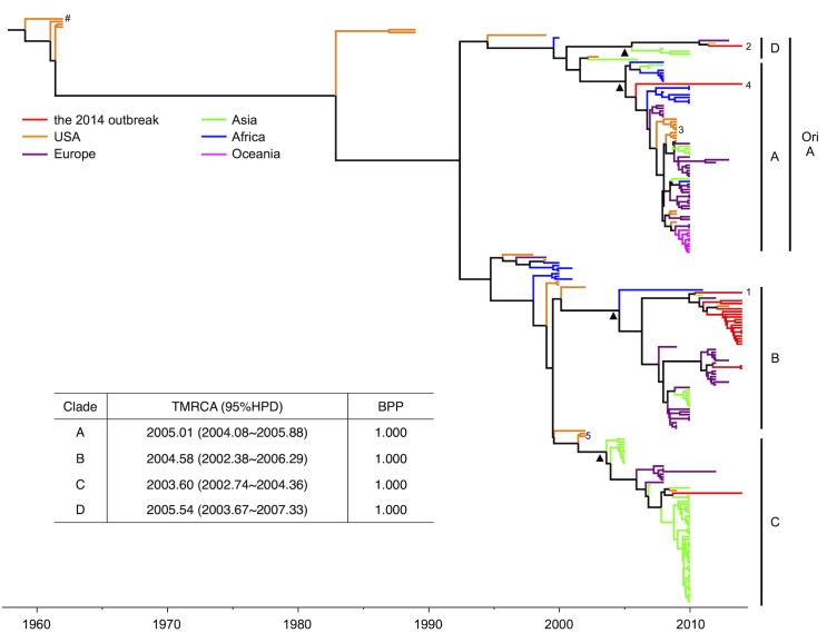
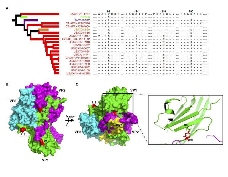
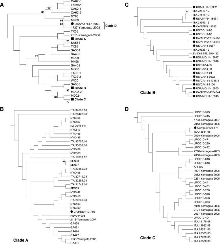
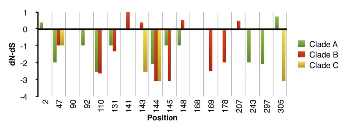
Enterovirus 68 (EVD68) causes respiratory illness, mostly in children. Despite a reported low-level of transmission, the occurrence of several recent outbreaks worldwide including the 2014 outbreak in North America has raised concerns regarding the pathogenesis and evolution of EVD68. To elucidate the phylogenetic features of EVD68 and possible causes for the 2014 outbreak, 216 EVD68 strain sequences were retrieved from Genbank, including 22 from the 2014 outbreak. Several geographic and genotypic origins were established for these 22 strains, 19 of which were classified as Clade B. Of these 19 strains, 17 exhibited subsequent clustering and variation in protein residues involved in host-receptor interaction and/or viral antigenicity. Approximately 18 inter-clade variations were detected in VP1, which led to the identification of a new Clade D in EVD68 strains. The classification of this new clade was also verified by the re-construction of a Neighbor-Joining tree during the phylogenetic analysis. In addition, our results indicate that members of Clade B containing highly specific alterations in VP1 protein residues were the foremost contributors to the 2014 outbreak in the US. Altered host-receptor interaction and/or host immune recognition may explain the evolution of EVD68 as well as the global emergence and ongoing adaptation of this virus.


Conflict of interest statement

Competing Interests: The authors have declared that no competing interests exist.

Figures

Fig 1
Fig 1. Bayesian MCMC analysis revealed Clade B members as the key contributors to the 2014 outbreak and demonstrated the emergence of a new Clade D from the original Clade.
A. Reported EVD68 sequences retrieved from GenBank are labeled in different colors in this tree based on sample origin, with the 2014 outbreak strains in red. The Bayesian phylogenetic tree was reconstructed with the partial VP1 coding region (2,435–3,128) according to a previously described method [26]. The original Clade A (ori A) classification is shown in addition to the four clades classified in this study. The scale bar below the tree indicates the time of strain sampling. Triangle markers indicate nodes for new clade classification. Specific strain labeling: Hashtag, EVD68 Fermon strain; 1, CA/AFP/11-1767; 2, US/KY/14-18953; 3, NYC strains; 4, CA/RESP/10-786; 5, MD02-1 and MD02-2. The year of the most recent common ancestor (TMRCA) and the Bayesian posterior probability (BPP) for each clade are shown in the table.
Fig 2
Fig 2. Specific residue variations detected in the EVD68 VP1 protein from the 2014 outbreak strains.
(A) VP1 protein alignment based on the Clade B subcluster. The alignment was performed with MEGA5 [36]. Hyphens indicate residues identical to those in CA/AFP/11-1767; “X” indicates unidentified amino acid due to unidentified nucleotides in the submitted CA/AFP/v12T04950 sequence. Numbers indicate protein positions. Sequence CQ5585 is missing the fragment surrounding position 290. The subtree for the subclustered Clade B strains is extracted from the tree in Fig 1. (B) Structures of viral proteins VP1 (green), VP2 (purple), and VP3 (cyan) in the surface model. The remodeling was based on the published EVD68 structure (PDB: 4WM8) [20]. Positions 218 and 290, located on the surface of VP1, are labeled in red. Position 99 is shown in red instead of 98 since the latter is missing in the published EVD68 structure. (C) Position 194, is located beneath the viral surface and is essential for β-sheet formation, which may be important for supporting EVD68 virion “canyon” formation. Left panel: the surface model of VP1 rotated 120° from the model shown in Fig 2B; right panel: a cartoon model of the β-sheet in which residue 194 interacts with residue 183.
Fig 3
Fig 3. Phylogenetic analysis of full-length VP1 region of EVD68 strains.
117 full-length EVD68 VP1 sequences were used for the re-construction of the Neighbor-Joining tree (A). Similar clade formation was detected compared to that in Fig 1, and each clade was shown as a subtree (B, C, and D, for Clade A, B, and C respectively). The tree was re-constructed with the help of MEGA5 [36]. Only bootstrap values >70 were shown. For a clearer presentation of the relationship between strains, only the topology was shown. Circle symbols indicate EVD68 strain from the 2014 outbreak, while square symbols indicate compressed EVD68 clades.
Fig 4
Fig 4. Evolutionary selection on the 18 clade-specific positions.
After sequence alignment, the dN-dS values for each clade were calculated for these clade-specific positions with the help of MEGA5 [36]. Values >0 indicate positive selection, while values <0 indicate purifying selection. Each codon with dN-dS = 0 in this study shared conserved nucleotides and thus was considered as purifyingly selected. Clade D was excluded from this analysis because only two strains contained full-length VP1.
Fig 5
Fig 5. Host-receptor binding and/or viral antigenicity correlate with EVD68 clade classification.
(A). Surface models of EVD68 VP1 (green), VP2 (purple), and VP3 (cyan), with clade-specific residues labeled in red. The remodeling was based on the published EVD68 structure (PDB: 4WM8) [20]. Positions 140 and 149 are labeled in red to indicate the approximate positions of residues 141, 143, 144, 145, and 148 that are missing in the published structure. (B). Structural alignment of VP1 from the published EVD68 structure (green) or the proposed structure (blue) based on the viral structure of Echovirus 7 (PDB: 2X5I) [38]. The alignment was performed with PyMol. (C). Surface model of the proposed EVD68 VP1 structure (blue), with clade-specific residues indicated in red. The panel on the right provides a detailed cartoon model showing clade-specific residues (in red) located on the loops forming the “canyon” responsible for host-receptor interaction. The BC-loop and DE-loop, thought to be critical in both host-receptor binding and viral antigenicity [40], are labeled with orange frames.

References

    1. Zhao K, Han X, Wang G, Hu W, Zhang W, Yu XF. Circulating coxsackievirus A16 identified as recombinant type A human enterovirus, China. Emerg Infect Dis. 2011;17(8):1537–1540. 10.3201/eid1708.101719 - DOI - PMC - PubMed
    1. McWilliam Leitch EC, Cabrerizo M, Cardosa J, Harvala H, Ivanova OE, Koike S, et al. The association of recombination events in the founding and emergence of subgenogroup evolutionary lineages of human enterovirus 71. J Virol. 2012;86(5):2676–2685. 10.1128/JVI.06065-11 - DOI - PMC - PubMed
    1. Wang X, Zhu C, Bao W, Zhao K, Niu J, Yu XF, et al. Characterization of full-length enterovirus 71 strains from severe and mild disease patients in northeastern China. PLoS One. 2012;7(3):e32405 10.1371/journal.pone.0032405 - DOI - PMC - PubMed
    1. Bessaud M, Razafindratsimandresy R, Nougairede A, Joffret ML, Deshpande JM, Dubot-Peres A, et al. Molecular comparison and evolutionary analyses of VP1 nucleotide sequences of new African human enterovirus 71 isolates reveal a wide genetic diversity. PLoS One. 2014;9(3):e90624 10.1371/journal.pone.0090624 - DOI - PMC - PubMed
    1. Li J, Chang J, Liu X, Yang J, Guo H, Wei W, et al. Protection from lethal challenge in a neonatal mouse model by circulating recombinant form coxsackievirus A16 vaccine candidates. J Gen Virol. 2014;95(Pt 5):1083–1093. 10.1099/vir.0.063560-0 - DOI - PMC - PubMed

Publication types

LinkOut - more resources