. 2015 Dec 2;10(12):e0143963.
doi: 10.1371/journal.pone.0143963. eCollection 2015.

Isolation, N-glycosylations and Function of a Hyaluronidase-Like Enzyme from the Venom of the Spider Cupiennius salei

Affiliations

Isolation, N-glycosylations and Function of a Hyaluronidase-Like Enzyme from the Venom of the Spider Cupiennius salei

Olivier Biner et al. PLoS One. .

Abstract

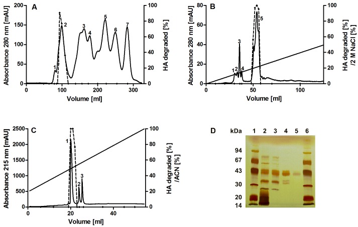
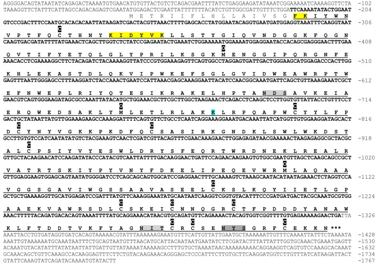
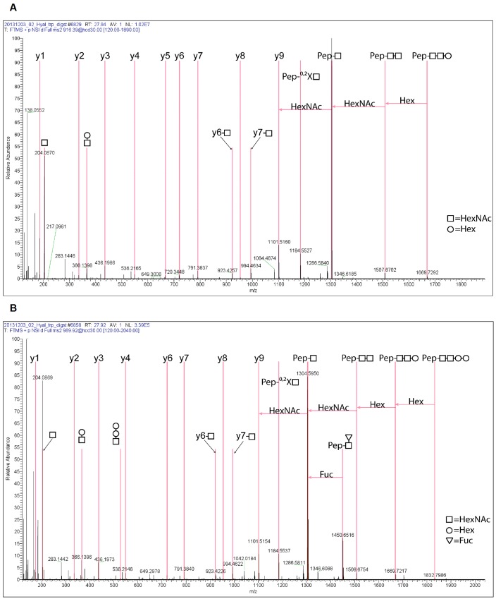
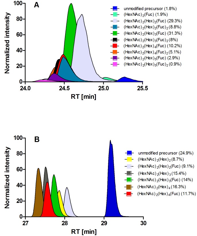
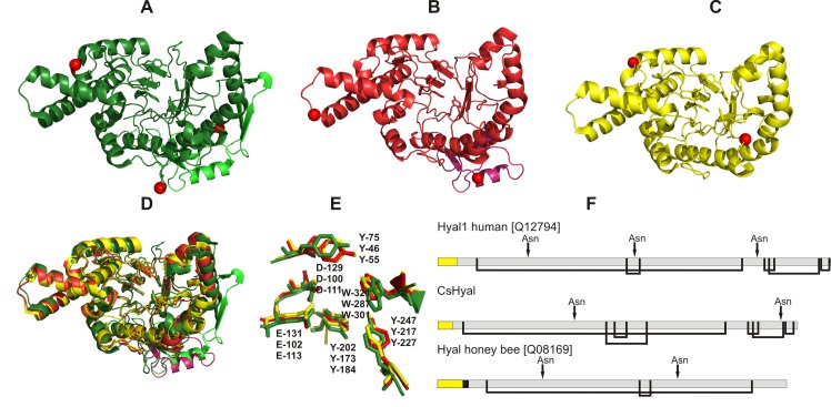
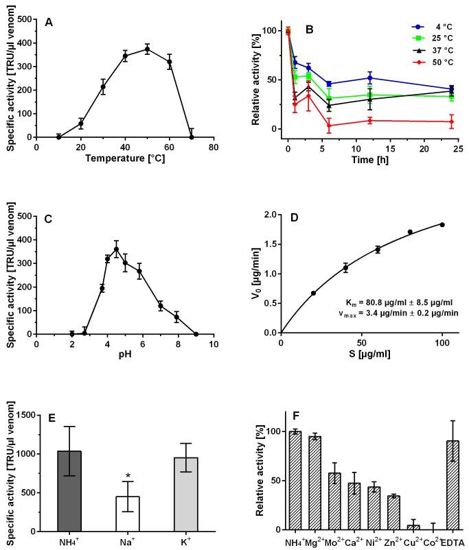
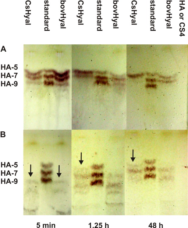
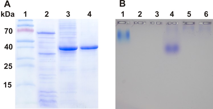
Structure of cupiennius salei venom hyaluronidase: Hyaluronidases are important venom components acting as spreading factor of toxic compounds. In several studies this spreading effect was tested on vertebrate tissue. However, data about the spreading activity on invertebrates, the main prey organisms of spiders, are lacking. Here, a hyaluronidase-like enzyme was isolated from the venom of the spider Cupiennius salei. The amino acid sequence of the enzyme was determined by cDNA analysis of the venom gland transcriptome and confirmed by protein analysis. Two complex N-linked glycans akin to honey bee hyaluronidase glycosylations, were identified by tandem mass spectrometry. A C-terminal EGF-like domain was identified in spider hyaluronidase using InterPro. The spider hyaluronidase-like enzyme showed maximal activity at acidic pH, between 40-60°C, and 0.2 M KCl. Divalent ions did not enhance HA degradation activity, indicating that they are not recruited for catalysis.

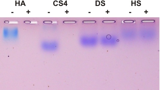
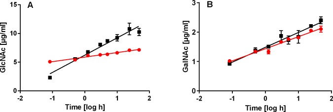
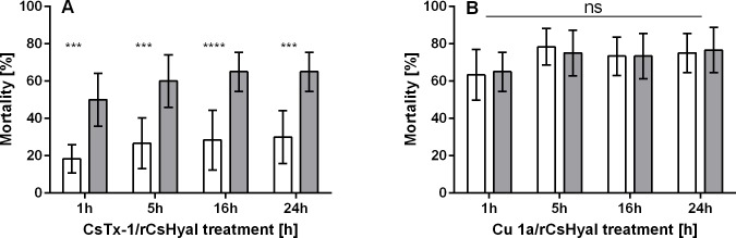
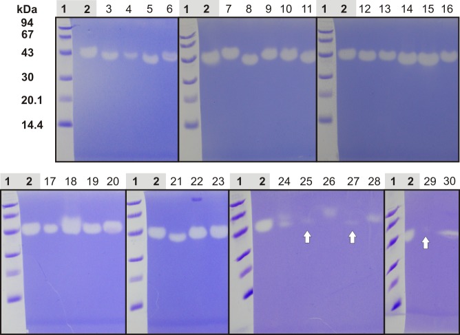
Function of venom hyaluronidases: Besides hyaluronan, the enzyme degrades chondroitin sulfate A, whereas heparan sulfate and dermatan sulfate are not affected. The end products of hyaluronan degradation are tetramers, whereas chondroitin sulfate A is mainly degraded to hexamers. Identification of terminal N-acetylglucosamine or N-acetylgalactosamine at the reducing end of the oligomers identified the enzyme as an endo-β-N-acetyl-D-hexosaminidase hydrolase. The spreading effect of the hyaluronidase-like enzyme on invertebrate tissue was studied by coinjection of the enzyme with the Cupiennius salei main neurotoxin CsTx-1 into Drosophila flies. The enzyme significantly enhances the neurotoxic activity of CsTx-1. Comparative substrate degradation tests with hyaluronan, chondroitin sulfate A, dermatan sulfate, and heparan sulfate with venoms from 39 spider species from 21 families identified some spider families (Atypidae, Eresidae, Araneidae and Nephilidae) without activity of hyaluronidase-like enzymes. This is interpreted as a loss of this enzyme and fits quite well the current phylogenetic idea on a more isolated position of these families and can perhaps be explained by specialized prey catching techniques.


Conflict of interest statement

Competing Interests: The authors have declared that no competing interests exist.

Figures

Fig 1
Fig 1. Purification of native CsHyal.
(A) Size exclusion chromatography of 400 μl crude venom from C. salei on a Bio-Gel P-60 column (2 x 100 cm, flow-rate: 0.7 ml/min) in 0.2 M NH4Ac buffer, pH 5.5. (B) Cation exchange chromatography of Hyal-active fractions on a MonoS column (HR 10/10, flow-rate: 2 ml/min) with 50 mM NH4Ac buffer, pH 5. Fractions were eluted in 60 min by applying a gradient of 0–50% of the same buffer containing 2 M NaCl. (C) RP-HPLC of Hyal-active fractions on a Nucleosil 300 butyl column (4.6 x 250 mm, 5 μm, flow-rate: 1 ml/min) equilibrated with 0.1% TFA (v/v) in water. Fractions were eluted in 80 min by applying a gradient of 20–100% solvent B 0.1% TFA (v/v) in ACN. (D) Purity of chromatographic fractions after the different separation steps was controlled by SDS PAGE (12.5%). Lanes: 1 and 6, low molecular mass markers (14.4–94 kDa, GE Healthcare); 2, crude venom; 3, gel filtration fraction 2; 4, MonoS fraction 5; 5, RP-HPLC fraction 1.
Fig 2
Fig 2. cDNA and amino acid sequence of CsHyal.
The deduced amino acid sequence is presented below the nucleotide sequence. The amino acid sequence of the mature protein is underlined and the corresponding nucleotide sequence is in bold. The gray doubly underlined sequences correspond to experimentally determined glycosylation sites and the dark-gray doubly underlined sequence to a tentative glycosylation site. The asterisks mark the stop codon. The sequence determined by tandem mass spectrometry results in a merged sequence coverage of the trypsin, GluC, and trypsin/LysC cleavage of purified CsHyal. The total sequence coverage including the signal peptide is 94.2%. Amino acids (not colored) have percolator scores with q < 0.01, those in blue are not trustworthy and not identified amino acid residues are colored in yellow. The letter C (black boxed) above the sequence stands for carboxyamidomethylcysteine and O (black boxed) for oxidation of methionine. The analysis of MS data sets was done with Proteome Discoverer Software (version 1.4.0.288, Thermo Fisher Scientific, CA, USA).
Fig 3
Fig 3. Comparison of the tandem mass spectra of a fucosylated and non-fucosylated glycopeptide originating from the glycosylation site at N360.
(A) Positive ESI fragment ion spectrum of the peptide FYAGNITCR modified with the N-glycan of the composition (HexNAc)2(Hex)2. The [M+2H]2+ precursor at 916.394 m/z was fragmented with HCD at a normalized collision energy of 30. The peptide y ion series as well as the relevant fragment ions for the identification of the N-glycan are indicated. (B) Positive ESI fragment ion spectrum of the peptide FYAGNITCR modified with the N-glycan of the composition (HexNAc)2(Hex)2(Fuc). The [M+2H]2+ precursor at 989.423 m/z was fragmented with HCD at a normalized collision energy of 30. The peptide y ion series as well as the relevant fragment ions for the identification of the N-glycan are indicated.
Fig 4
Fig 4. Semi-quantitative extraction of glycopeptide abundances by MS 1 filtering.
The precursor ion mass of the identified glycopeptides was used to extract the chromatographic area. The percental distribution of the abundances of the different N-glycans within the two glycosylation sites was calculated. Plot y-axis were normalized to the most abundant value in the graph. N-glycans exhibiting a normalized area of < 2% are not shown in this plot. (A) Area plot of the different glycopeptides on glycosylation site N134. (B) Area plot of the different glycopeptide subforms on glycosylation site N360.
Fig 5
Fig 5. Structural comparison of different Hyals.
The residues of the active sites are shown as sticks and experimentally determined N-linked glycosylation sites are indicated by red spheres (A-C). The Hyal domain is colored in green and the EGF-like domain in bright green (A, human Hyal-1), the Hyal domain in red and the EGF-like domain in pink (B, C. salei), and the Hyal domain in yellow (C, bee venom Hyal). All structures were drawn using PyMOL. (A) Crystal structure of human Hyal-1 (PDB:2PE4) determined by Chao et al. [48]. (B) Structure of CsHyal modeled using the human Hyal-1 structure as template. (C) Crystal structure of bee venom Hyal (PDB:1FCQ) determined by Markovic-Housley et al. [49]. (D) Overlay/alignment of the structures of human Hyal-1 (green), C. salei Hyal (red) and bee venom Hyal (yellow). Active site residues are shown as sticks and EGF-like domains are colored in green and pink, respectively. (E) Zoom of the structures in D shows the active site residues. (F) Disulfide bridge arrangement and glycosylation sites of human, C. salei and honey bee Hyal. Yellow boxes refer to signal peptides, black box to propeptide and gray boxes to mature proteins.
Fig 6
Fig 6. Enzymatic characterization of native CsHyal.
(A) Effect of temperature on CsHyal activity. (B) Thermostability of CsHyal measured at 4, 25, 37, and 50°C after 1, 3, 6, 12, and 24 h of incubation. (C) pH profile of CsHyal activity. (D) Determination of Km and Vmax using non-linear regression. (E) Effect of monovalent salts in 0.2 M NH4 +-buffer containing 0.2 M K+ or Na+, respectively. Activity decreased in Na+ compared to NH4 + (* significance, p = 0.007). (F) Influence of divalent cations and EDTA on CsHyal activity.
Fig 7
Fig 7. Substrate specificity of native CsHyal.
HA, CS4, HS, and DS were incubated with 0.1 μl venom at 40°C overnight and separated on a 1% agarose gel at 50 mA for 3h. Degradation was revealed by sequential toluidine blue and Stains-All staining. Lanes labeled with + represent incubation in the presence of venom and those with - in the absence of venom.
Fig 8
Fig 8. Time dependent production of terminal GlcNAc or GalNAc residues at the reducing end through C. salei venom and bovHyal.
(A) Incubation of HA with C. salei venom (red circles) or bovHyal (black squares) resulted in a time dependent release of reducing GlcNAc as indirect measurement of Hyal activity. (B) Incubation of CS4 with C. salei venom (red circles) or bovHyal (black squares) resulted in a time dependent release of reducing GalNAc as indirect measurement of Hyal activity.
Fig 9
Fig 9. TLC of time dependent degradation products of HA and CS4 through hydrolysis with C. salei venom or bovHyal.
(A) HA was incubated with C. salei venom or bovHyal for 5 min, 1.25 h and 48 h. (B) CS4 was incubated with C. salei venom or bovHyal for 5 min, 1.25 h and, 48 h as described under materials and methods. Standards were different HA-oligomers with an additional GlcNAc at the non-reducing end: HA-5, HA-7 and, HA-9. HA or CS4 without C. salei venom/bovHyal were applied as control.
Fig 10
Fig 10. Purification of rCsHyal and substrate specificity of purified rCsHyal.
(A) Solubility of expressed rCsHyal and the purity of separated and refolded rCsHyal were analyzed by SDS PAGE (12%). rCsHyal was only found in the insoluble fraction and was therefore purified under denaturing conditions. Lanes: 1, PageRuler Prestained Protein Ladder (Thermo Scientific); 2, soluble fraction after cell lysis; 3, insoluble fraction after cell lysis; 4, rCsHyal after purification and refolding. (B) HA (lane 1, 2 and 3) and CS4 (lane 4, 5 and 6) were incubated with approximately 1.52 TRU (lane 2 and 5) and 0.76 TRU (lane 3 and 6) rCsHyal at 40°C overnight and separated on a 1% agarose gel at 50 mA for 3h. Degradation was revealed by sequential toluidine blue and Stains-All staining.
Fig 11
Fig 11. Bioassays with D. melanogaster.
Not shown are control injections of only buffer or rCsHyal (0.42 TRU/mg fly) which were completely non-toxic. (A) The neurotoxin CsTx-1 (0.40 pmol/mg fly) was injected alone (open bars) or in combination with rCsHyal (0.42 TRU/mg fly) (gray bars) and the total paralysis/mortality was determined after 1, 5, 16, and 24h. **** corresponds to p < 0.0001 and *** corresponds to p < 0.001. (B) The cytolytic acting Cu 1a (7.45 pmol/mg fly) was injected alone (open bars) or in combination with rCsHyal (0.42 TRU/mg fly) (gray bars) and the total paralysis/mortality was determined after 1, 5, 16, and 24h. ns means not significant.
Fig 12
Fig 12. Hyaluronidase activity of different spider venoms.
Different spider venoms were separated in a 10% SDS-PAGE which was copolymerized with HA as substrate for Hyals. The dilution of different venoms is given in brackets. Lanes: 1, low molecular mass standard (GE Healthcare); 2, C. salei (1:100); 3, Phoneutria fera (1:20); 4, Phoneutria reidyi (1:10); 5, Eusparassus dufouri (1:10); 6, Uroctea durandi (1:10); 7, Ancylometes rufus (1:100); 8, Viridasius fasciatus (1:50); 9, Hogna radiata (1:100); 10, Alopecosa fabrilis (1:100); 11, Lycosa praegrandis (1:100); 12, Dolomedes okefinokensis (1:100); 13, Zoropsis spinimana (1:100); 14, Polybetes pythagoricus (1:100); 15, Isopeda villosa (1:100); 16, Lycosa hispanica (1:100); 17, Geolycosa vultuosa (1:10); 18, Pisaura mirabilis (1:20); 19, Macrothele calpeiana (1:20); 20, Meta menardi (1:10); 21, Latrodectus tredecimguttatus (1:100); 22, Sericopelma rubronitens (1:100); 23, Drassodes lapidosus (1:10); 24, Steatoda paykulliana (1:5); 25, Alopecosa marikovskyi (1:5); 26, Linothele megatheloides (1:5); 27, Oxyopes sp. (1:5); 28, Segestria florentina (1:5); 29, Filistata insidiatrix (1:5); and 30, Tegenaria atrica (1:20).

Similar articles

Cited by

References

    1. Stern R, Jedrzejas MJ. Hyaluronidases: their genomics, structures, and mechanisms of action. Chem Rev. 2006;106(3):818–839. - PMC - PubMed
    1. Stern R, Kogan G, Jedrzejas MJ, Šoltés L. The many ways to cleave hyaluronan. Biotechnol Adv. 2007;25(6):537–557. - PubMed
    1. Sugahara K, Mikami T, Uyama T, Mizuguchi S, Nomura K, Kitagawa H. Recent advances in the structural biology of chondroitin sulfate and dermatan sulfate. Curr Opin Struct Biol. 2003;13(5):612–620. - PubMed
    1. Feng L, Gao R, Gopalakrishnakone P. Isolation and characterization of a hyaluronidase from the venom of Chinese red scorpion Buthus martensi . Comp Biochem Physiol Toxicol Pharmacol. 2008;148(3):250–257. - PubMed
    1. Kemeny DM, Dalton N, Lawrence AJ, Pearce FL, Vernon CA. The purification and characterisation of hyaluronidase from the venom of the honey bee, Apis mellifera . Eur J Biochem. 1984;139(2):217–223. - PubMed

MeSH terms