. 2015 Dec 2:14:490.
doi: 10.1186/s12936-015-1014-7.

Transfection with thymidine kinase permits bromodeoxyuridine labelling of DNA replication in the human malaria parasite Plasmodium falciparum

Affiliations

Transfection with thymidine kinase permits bromodeoxyuridine labelling of DNA replication in the human malaria parasite Plasmodium falciparum

Catherine J Merrick. Malar J. .

Abstract

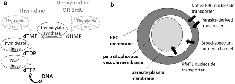
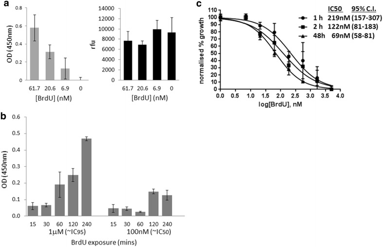
Background: Plasmodium falciparum, the causative agent of severe human malaria, is an early-diverging protozoan whose lifecycle has many unusual features, including its modes of replication. Research on the Plasmodium cell cycle, which occurs primarily via schizogony instead of canonical binary fission, has been hampered by a lack of tools and markers that can be transferred from cell cycle studies in model organisms. A common tool used to study DNA replication and the cell cycle in human cells is the labelling of newly-replicated DNA with the modified nucleotide bromodeoxyuridine (BrdU), followed by immunofluorescent detection. Plasmodium parasites, however, do not incorporate BrdU because they rely only on de novo synthesis of pyrimidines and do not salvage thymidine analogues like BrdU for conversion into nucleotides.

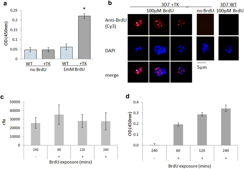
Methods: Analysis of biochemical pathways in Plasmodium indicated that the absence of the enzyme thymidine kinase (TK) may be the only impediment to BrdU incorporation in this organism. A TK gene from Herpes simplex was, therefore, introduced into the Plasmodium falciparum 3D7 strain and the effect on BrdU labelling was assessed by enzyme-linked immunosorbent assay and immunofluorescence microscopy.

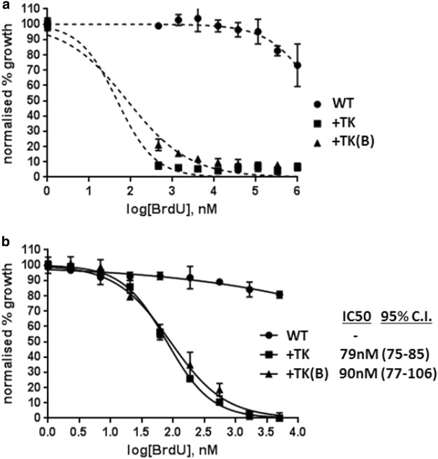
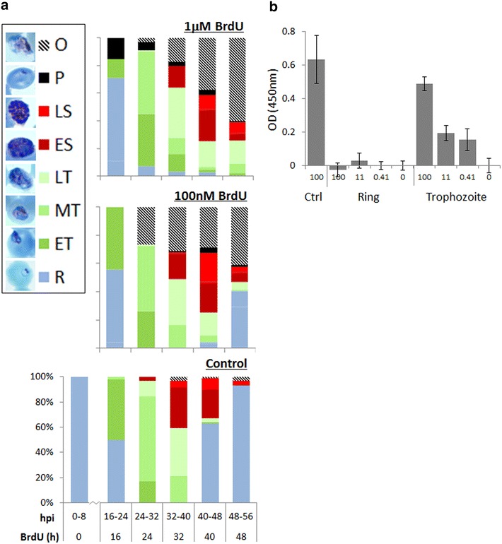
Results: Introduction of a TK gene produces parasites that can indeed incorporate BrdU. This forms a sensitive indicator of DNA replication, which can be detected by both quantitative and qualitative assays on either a population level or a single-cell level. Plasmodium falciparum, when expressing TK, becomes unusually sensitive to BrdU toxicity.

Conclusions: BrdU labelling represents a significant new tool for investigating DNA replication and the cell cycle in Plasmodium.


Figures

Fig. 1
Fig. 1
Schematic of nucleotide metabolism and transport pathways in P. falciparum. a Enzymes required for the metabolism of pyrimidine nucleosides to nucleotides for incorporation into DNA. Plasmodium species lack thymidine kinase but contain all the other enzymes shown downstream. b Schematic showing the route by which modified nucleosides like BrdU could enter intra-erythrocytic parasites. RBC red blood cell, PfNT1 P. falciparum nucleoside transporter 1
Fig. 2
Fig. 2
TK-expressing P. falciparum can incorporate BrdU, which is a sensitive indicator of DNA replication. a ELISA on wild-type (WT) and +TK parasites (2 × 105 parasites/well) after exposure to 1 mM BrdU for 4 h at the trophozoite stage. Error bars show standard deviation of triplicate readings; star indicates the only significantly differing dataset (One-way ANOVA, p < 0.0001). b Immunofluorescence assay on (WT) and +TK parasites exposed to 100 mM BrdU for 12 h at trophozoite stage. c SYBR Green I staining of +TK parasites after exposure to no BrdU (−) or to 100 mM BrdU (+) for 1, 2 or 4 h. Parasites were released from erythrocytes and suspended in PBS [as for the ELISA assay shown in (a)], then plated with MSF lysis buffer at 1 × 106 parasites/well. The increase in DNA content over 3 h is not detectable by this MSF-type assay. Error bars show standard deviation of triplicate readings. rfu, relative fluorescence units. d ELISA on the same +TK parasites as in (c), plated at 1x10⁶ parasites/well after exposure to no BrdU (−) or to 100 mM BrdU (+) for 1, 2 or 4 h. Background signal, obtained from the same parasites incubated without BrdU, has been subtracted from all readings, and error bars show standard deviation of triplicate readings. Figure c confirms that approximately the same numbers of parasites were plated in all cases
Fig. 3
Fig. 3
TK-expressing parasites become sensitive to BrdU toxicity. a MSF assay on WT and two different TK-expressing parasite lines, +TK and +TK(B), over a BrdU range of 1–0.46 mM. Error bars show standard deviation of triplicate readings. b MSF assay on the same parasites as in (a), over a range of 5–2.3 nM BrdU. IC50 values were calculated with GraphPad Prism software, 95 % confidence intervals are given in parentheses
Fig. 4
Fig. 4
BrdU causes schizont-stage arrest in TK-expressing parasites. a Developmental stages of +TK parasites assessed at 8 h intervals over a single growth cycle in the presence of 0, 100 nM or 1 mM BrdU. 100 parasites were counted at each timepoint. Photographs show representative parasite morphology at each stage: R ring, ET early trophozoite (parasite less than half the width of host cell), MT middle trophozoite (parasite more than half the width of, but not entirely filling, host cell), LT late trophozoite (parasite filling all or nearly all of host cell), ES early schizont (nuclear masses visible within parasite), LS late schizont (defined merozoites visible), P pyknotic (parasite stained as a dense shrunken intracellular dot without clear morphological features), O parasite outside erythrocyte, hpi hours post invasion. b ELISA on +TK parasites, plated at 1 × 106 parasites/well, after ring-stage exposure to 0, 0.41, 11 or 100 mM BrdU for 4 h. Parasites were either harvested immediately after washing out the BrdU, as rings, or cultured until the late-trophozoite stage and then harvested for ELISA. Control (ctrl) parasites were cultured to the trophozoite stage before exposing to 100 mM BrdU for 4 h at this stage and then harvesting immediately. The background signal, obtained from parallel samples incubated without BrdU, has been subtracted from each set of readings. Error bars show standard deviation of triplicate readings
Fig. 5
Fig. 5
Toxicity does not preclude the use of BrdU as a short-term labelling agent in P. falciparum. a ELISA on +TK parasites, plated at 1 × 106 parasites/well, after 48 h exposure to the three lowest concentrations of BrdU used in the MSF assay shown in Fig. 3b. Background signal, obtained from the sample incubated without BrdU, has been subtracted from all readings. Error bars show standard deviation of triplicate readings. A parallel MSF-type assay on the same parasites shows that DNA content is similar in all samples, confirming that within 48 h, severe growth inhibition has not occurred at these sub-IC50 exposures. b ELISA on +TK parasites (1 × 106 parasites/well), after pulses of BrdU from 15 min to 4 h, applied at the trophozoite stage. The background signal, obtained from parallel samples incubated without BrdU, has been subtracted from each set of readings. Error bars show standard deviation of triplicate readings. c MSF assay on +TK parasites after 1 or 2 h pulses of BrdU (5 mM to 2.3 nM) at trophozoite stage, followed by washing and growth in fresh medium for 48 h. Parasites were also kept in BrdU for the full 48 h, as in Fig. 3b, for comparison. Error bars show standard deviation of triplicate readings. IC50 values were calculated with GraphPad Prism software, 95 % confidence intervals are given in parentheses

References

    1. WHO: World Malaria Report 2014. Geneva, World Health Organization, 2014.
    1. Arnot DE, Gull K. The Plasmodium cell-cycle: facts and questions. Ann Trop Med Parasitol. 1998;92:361–365. doi: 10.1080/00034989859357. - DOI - PubMed
    1. Arnot DE, Ronander E, Bengtsson DC. The progression of the intra-erythrocytic cell cycle of Plasmodium falciparum and the role of the centriolar plaques in asynchronous mitotic division during schizogony. Int J Parasitol. 2011;41:71–80. doi: 10.1016/j.ijpara.2010.07.012. - DOI - PubMed
    1. Gerald N, Mahajan B, Kumar S. Mitosis in the human malaria parasite Plasmodium falciparum. Eukaryot Cell. 2011;10:474–482. doi: 10.1128/EC.00314-10. - DOI - PMC - PubMed
    1. Doerig C, Endicott J, Chakrabarti D. Cyclin-dependent kinase homologues of Plasmodium falciparum. Int J Parasitol. 2002;32:1575–1585. doi: 10.1016/S0020-7519(02)00186-8. - DOI - PubMed

Publication types

LinkOut - more resources