. 2015 Dec 2:12:110.
doi: 10.1186/s12984-015-0102-9.

Improving robustness against electrode shift of high density EMG for myoelectric control through common spatial patterns

Affiliations

Improving robustness against electrode shift of high density EMG for myoelectric control through common spatial patterns

Lizhi Pan et al. J Neuroeng Rehabil. .

Abstract

Background: Most prosthetic myoelectric control studies have concentrated on low density (less than 16 electrodes, LD) electromyography (EMG) signals, due to its better clinical applicability and low computation complexity compared with high density (more than 16 electrodes, HD) EMG signals. Since HD EMG electrodes have been developed more conveniently to wear with respect to the previous versions recently, HD EMG signals become an alternative for myoelectric prostheses. The electrode shift, which may occur during repositioning or donning/doffing of the prosthetic socket, is one of the main reasons for degradation in classification accuracy (CA).

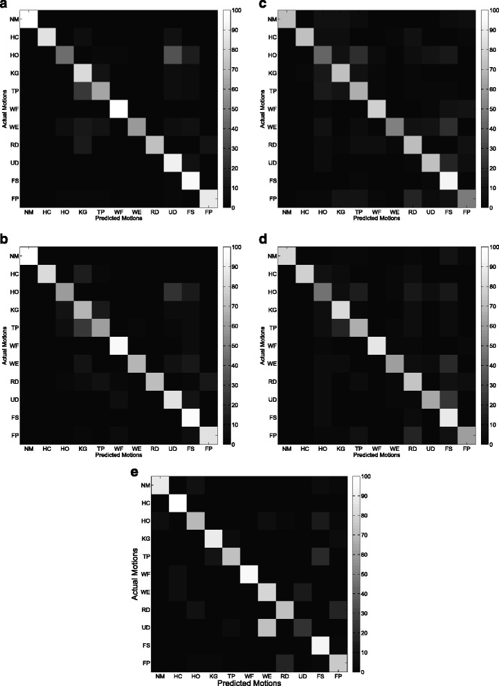
Methods: HD EMG signals acquired from the forearm of the subjects were used for pattern recognition-based myoelectric control in this study. Multiclass common spatial patterns (CSP) with two types of schemes, namely one versus one (CSP-OvO) and one versus rest (CSP-OvR), were used for feature extraction to improve the robustness against electrode shift for myoelectric control. Shift transversal (ST1 and ST2) and longitudinal (SL1 and SL2) to the direction of the muscle fibers were taken into consideration. We tested nine intact-limb subjects for eleven hand and wrist motions. The CSP features (CSP-OvO and CSP-OvR) were compared with three commonly used features, namely time-domain (TD) features, time-domain autoregressive (TDAR) features and variogram (Variog) features.

Results: Compared with the TD features, the CSP features significantly improved the CA over 10 % in all shift configurations (ST1, ST2, SL1 and SL2). Compared with the TDAR features, a. the CSP-OvO feature significantly improved the average CA over 5 % in all shift configurations; b. the CSP-OvR feature significantly improved the average CA in shift configurations ST1, SL1 and SL2. Compared with the Variog features, the CSP features significantly improved the average CA in longitudinal shift configurations (SL1 and SL2).

Conclusion: The results demonstrated that the CSP features significantly improved the robustness against electrode shift for myoelectric control with respect to the commonly used features.


Figures

Fig. 1
Fig. 1
Position of the HD EMG grid and the HD EMG grid of 192 electrodes used in the experiments
Fig. 2
Fig. 2
Shift transversal to the direction of the muscle fibers. Shift leftwards (ST1): the white color electrodes were used for training, while the red color electrodes were used for testing. Shift rightwards (ST2): the red color electrodes were used for training, while the white color electrodes were used for testing
Fig. 3
Fig. 3
Shift longitudinal to the direction of the muscle fibers. Shift downwards (SL1): the white color electrodes were used for training, while the red color electrodes were used for testing. Shift upwards (SL2): the red color electrodes were used for training, while the white color electrodes were used for testing
Fig. 4
Fig. 4
Last CSP pattern of motion 1 and motion 4 for CSP-OvO extension scheme in transversal direction shift (ST1). Left and right columns were the CSP patterns before and after electrode shift
Fig. 5
Fig. 5
Last CSP pattern of motion 1 and motion 4 for CSP-OvO extension scheme in longitudinal direction shift (SL1). Left and right columns were the CSP patterns before and after electrode shift
Fig. 6
Fig. 6
First CSP pattern of each active motion and rest motions for CSP-OvR extension scheme in transversal direction shift (ST1). First and second columns were the CSP patterns of the first five active motions (HC, HO, KG, TP and WF) before and after electrode shift. Third and fourth columns were the CSP patterns of the last five active motions (WE, RD, UD, FS and FP) before and after electrode shift
Fig. 7
Fig. 7
First CSP pattern of each active motion and rest motions for CSP-OvR extension scheme in longitudinal direction shift (SL1). Left and right columns were the CSP patterns before and after electrode shift
Fig. 8
Fig. 8
Average CA across all subjects for five features: CSP-OvO, CSP-OvR, TD, TDAR and Variog. Error bars represented the standard deviation. The tests were marked by * in which significance were found between different features
Fig. 9
Fig. 9
Average confusion matrix of the five features across all subjects in ST1. a CSP-OvO. b CSP-OvR. c TD. d TDAR. e Variog
Fig. 10
Fig. 10
Average confusion matrix of the five features across all subjects in ST2. a CSP-OvO. b CSP-OvR. c TD. d TDAR. e Variog
Fig. 11
Fig. 11
Average confusion matrix of the five features across all subjects in SL1. a CSP-OvO. b CSP-OvR. c TD. d TDAR. e Variog
Fig. 12
Fig. 12
Average confusion matrix of the five features across all subjects in SL2. a CSP-OvO. b CSP-OvR. c TD. d TDAR. e Variog
Fig. 13
Fig. 13
Average RCS across all subjects for four features: CSP-OvO, CSP-OvR, TD, and TDAR. Error bars represented the standard deviation

References

    1. Farina D, Jiang N, Rehbaum H, Holobar A, Graimann B, Dietl H, et al. The extraction of neural information from the surface EMG for the control of upper-limb prostheses: emerging avenues and challenges. IEEE Trans. Neural Syst Rehabil Eng. 2014;22(4):797–809. doi: 10.1109/TNSRE.2014.2305111. - DOI - PubMed
    1. Graupe D, Cline WK. Functional separation of EMG signals via ARMA identification methods for prosthesis control purposes. IEEE Trans Syst Man Cybern. 1975;SMC-5(2):252–9. doi: 10.1109/TSMC.1975.5408479. - DOI
    1. Doerschuk PC, Gustafon DE, Willsky A. Upper extremity limb function discrimination using EMG signal analysis. IEEE Trans. Biomed. Eng. 1983;BME-30(1):18–29. doi: 10.1109/TBME.1983.325162. - DOI - PubMed
    1. Fougner A, Stavdahl O, Kyberd P, Losier Y, Parker P. Control of upper limb prostheses: Terminology and proportional myoelectric control-a review. IEEE Trans Neural Syst Rehabil Eng. 2012;20(5):663–77. doi: 10.1109/TNSRE.2012.2196711. - DOI - PubMed
    1. Hudgins B, Parker P, Scott RN. A new strategy for multifunction myoelectric control. IEEE Trans Biomed Eng. 1993;40(1):82–94. doi: 10.1109/10.204774. - DOI - PubMed

Publication types

LinkOut - more resources