. 2016 Aug 15:159:83-89.
doi: 10.1016/j.lfs.2015.11.026. Epub 2015 Nov 26.

Linagliptin attenuates diabetes-induced cerebral pathological neovascularization in a blood glucose-independent manner: Potential role of ET-1

Affiliations

Linagliptin attenuates diabetes-induced cerebral pathological neovascularization in a blood glucose-independent manner: Potential role of ET-1

Mohammed Abdelsaid et al. Life Sci. .

Abstract

Aims: We have shown that glycemic control with metformin or endothelin-1 (ET-1) inhibition with bosentan prevents and restores diabetes-mediated cerebral pathological remodeling and neovascularization. Our recent data suggest that linagliptin, a member of the dipeptidyl peptidase-4 inhibitor class of glucose-lowering agents, prevents cerebrovascular remodeling and dysfunction independent of its blood glucose lowering effects. We hypothesized that linagliptin prevents pathological neovascularization via the modulation of the ET-1 system.

Materials and methods: 24-week old diabetic Goto-Kakizaki and nondiabetic Wistar rats were treated for 4weeks with either vehicle chow or chow containing 166mg/kg linagliptin. At termination, FITC-dextran was injected to visualize the vasculature. Brain sections were imaged by confocal microscopy for vascular density, tortuosity, vascular volume, and surface in both the cortex and striatum. Retinal acellular capillary formation was measured. Brain microvascular endothelial cells (BMVEC) isolated from control or diabetic rats were treated with linagliptin with or without ET-1 dual receptor antagonist and tested for angiogenic properties with cell migration and tube formation assays.

Key finding: Linagliptin reduced all indices of cerebral neovascularization compared with control rats. In vitro, linagliptin normalized the augmented angiogenic properties of BMVECs isolated from diabetic animals and bosentan reversed this response. Cells from diabetic animals had higher ET-1 and less ETB receptors than in control cells. Linagliptin significantly decreased ET-1 levels and increased ETB receptors.

Significance: ET system contributes to pathological neovascularization in diabetes as evidenced by restoration of functional angiogenesis by bosentan treatment and prevention of linagliptin-mediated improvement of angiogenesis in the in vitro model.

Keywords: Diabetes; Endothelin; Linagliptin and neovascularization.


Conflict of interest statement

statement Authors declare no conflict of interest.

Figures
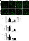
Fig. 1
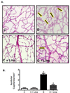
Fig. 1. Linagliptin decreases pathological neovascularization indices in diabetes
24 week old nondiabetic Wistar (C) and diabetic GK rats (D) were treated for 4 weeks with linagliptin 166 mg/kg chow (C+Lina, D+Lina). At termination, rats were injected FITC-dextran to fill and visualize blood vessels. Images acquired by confocal microscopy were used for 3-dimensional reconstruction of the cerebrovascular network for measurement of vascular density, vascular volume, and surface area in the cortex and striatum regions using Volocity 6 software. Representative images from cortex (upper panel) and striatum (lower panel) are shown on Panel (A) and average data are shown in histograms on Panels (B–D). Linagliptin treatment significantly reduced vascular volume, vascular density and surface area as compared to vehicle-treated diabetic GK rats (Results are expressed as mean ± SEM, n=3–5, *p<0.05 vs C, #p<0.05 vs D).
Fig. 2
Fig. 2. Linagliptin decreases small vessels remodeling in diabetes
Confocal images used for vascular density measurements in Fig. 1 were converted to binary mode and skeletonized using Image J software to determine branch density and tortuosity in the cortex area. (A) Representative binary (top panel) and skeleton images (bottom panels) of brain sections of Wistar and GK rats treated with vehicle or linagliptin. Linagliptin treatment significantly reduced tortuosity (B) and branch density (C) compared with vehicle-treated GK rats (Results are expressed as mean ± SEM, n=3–5, *p<0.05 vs C, #p<0.05 vs D)
Fig. 3
Fig. 3. Linagliptin decreases retinal acellular capillaries in diabetes
Retinal acellular capillary formation is a surrogate marker for pathological neovascularization in diabetes. 24 week old nondiabetic Wistar (C) and diabetic GK rats (D) were treated for 4 weeks with linagliptin 166mg/kg chow (C+Lina, D+Lina) and capillary-like structures that contain only basement membrane were counted (arrows) (n=4–5, *p<0.05 vs C, #p<0.05 vs D).
Fig. 4
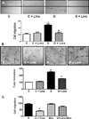
Fig. 4. Linagliptin restores augmented endothelial cell angiogenic properties in diabetes
BMVECs from diabetic animals (D) showed augmented angiogenic signaling as shown by increased tube formation compared to endothelial cells from Wistar control group (C). (A) Treatment with Linagliptin (100 nM) significantly decreased endothelial cell migration of GK rats (n=3–4, *p<0.05 vs C, #p<0.05 vs D). (B) In parallel, treatment with Linagliptin (100 nM) significantly decreased tube formation of GK BMVECs (n=3–4, *p<0.05 vs C, #p<0.05 vs D). (C) BMVECs from diabetic animals treated with vehicle (D), Linagliptin (100 nM) (D+Lina), Bosentan (10 µM) (Bos) or both linagliptin and bosentan (Bosentan was applied 15 min before linagliptin) (D+Lina+Bos). While treatment with bosentan alone did not impact the migratory properties of BMVECs from diabetic animals, it negated the inhibitory effect of linagliptin. (n=3, *p<0.05 vs D).
Fig. 5
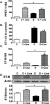
Fig. 5. Linagliptin modulates the ET system in diabetes
(A) PPET-1 transcription was assessed in control (C) and diabetic (D) BMVECs. Treatment with linagliptin showed significant increase in the PPET-1 mRNA levels (n=3, *p<0.05 vs C). (B) While BMVECs from diabetic animals showed 4-fold higher levels of ET-1 in the media, treatment with linagliptin reduced ET-1 levels. (n=3, *p<0.05 vs C, #p<0.05 vs D). (C–D) BMVECs from diabetic animals showed lower ETB mRNA and protein levels as compared to control cells. Treatment of control (C+Lina) and diabetic (D+Lina) BMVECs with linagliptin showed significant increase in the ETB mRNA and protein levels (n=3, *p<0.05 vs C, #p<0.05 vs GK).

References

    1. American Diabetes Association. National diabetes fact sheet. 2015 http://www.diabetes.org/diabetes-statistics.jsp.
    1. Ergul A, Abdelsaid M, Fouda AY, Fagan SC. Cerebral neovascularization in diabetes: implications for stroke recovery and beyond. J Cereb Blood Flow Metab. 2014;34:553–563. - PMC - PubMed
    1. Ergul A, Kelly-Cobbs A, Abdalla M, Fagan SC. Cerebrovascular complications of diabetes: Focus on stroke. Endocr Metab Immune Disord Drug Targets. 2012;12:148–158. - PMC - PubMed
    1. Li W, Prakash R, Kelly-Cobbs AI, Ogbi S, Kozak A, El-Remessy AB, Schreihofer DA, Fagan SC, Ergul A. Adaptive cerebral neovascularization in a model of Type 2 diabetes: Relevance to focal cerebral ischemia. Diabetes. 2010;59:228–235. - PMC - PubMed
    1. Prakash R, Johnson M, Fagan SC, Ergul A. Cerebral Neovascularization and Remodeling Patterns in Two Different Models of Type 2 Diabetes. PloS One. 2013;8:e56264. - PMC - PubMed