. 2016 Feb 1;27(3):491-9.
doi: 10.1091/mbc.E15-03-0161. Epub 2015 Dec 2.

A role of the sphingosine-1-phosphate (S1P)-S1P receptor 2 pathway in epithelial defense against cancer (EDAC)

Affiliations

A role of the sphingosine-1-phosphate (S1P)-S1P receptor 2 pathway in epithelial defense against cancer (EDAC)

Sayaka Yamamoto et al. Mol Biol Cell. .

Abstract

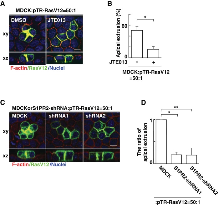
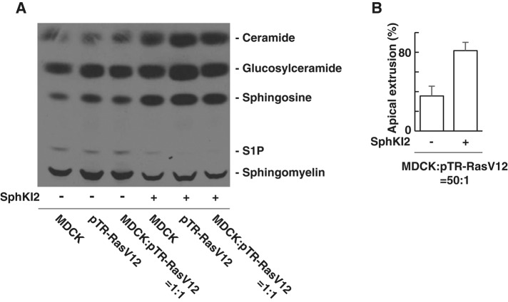
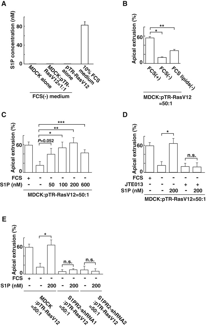
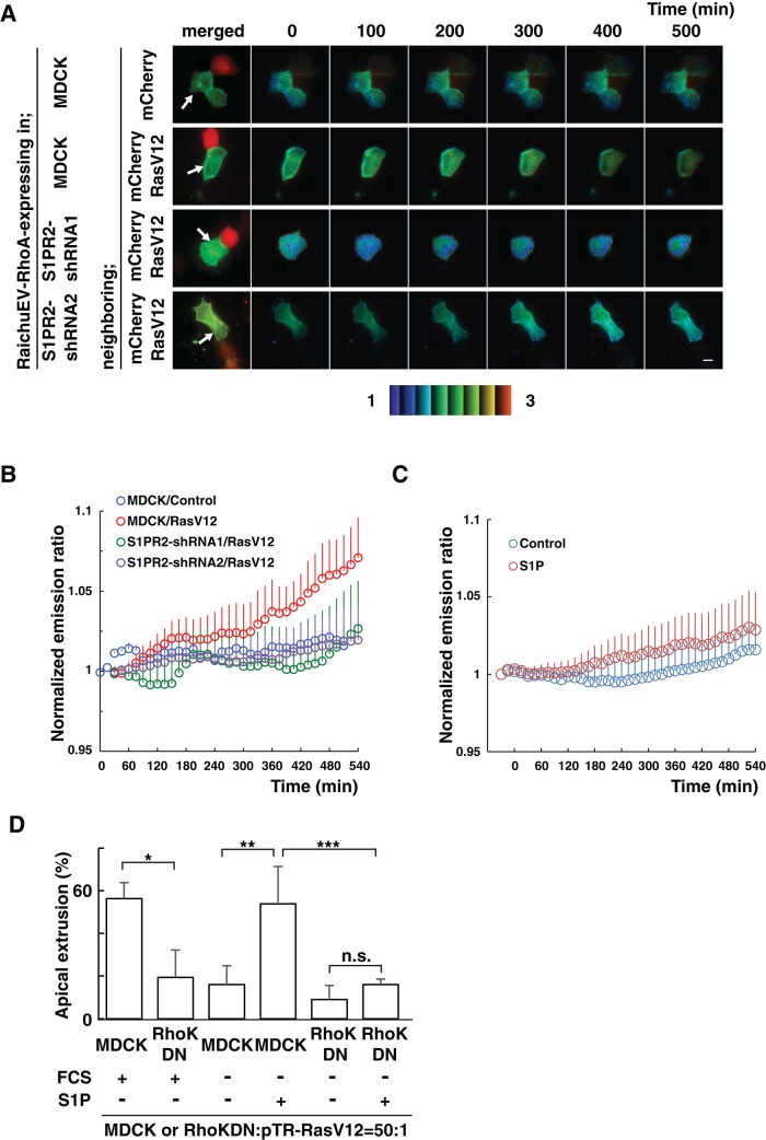
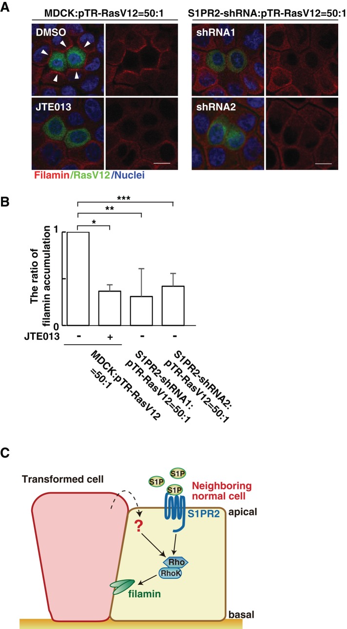
At the initial step of carcinogenesis, transformation occurs in single cells within epithelia, where the newly emerging transformed cells are surrounded by normal epithelial cells. A recent study revealed that normal epithelial cells have an ability to sense and actively eliminate the neighboring transformed cells, a process named epithelial defense against cancer (EDAC). However, the molecular mechanism of this tumor-suppressive activity is largely unknown. In this study, we investigated a role for the sphingosine-1-phosphate (S1P)-S1P receptor 2 (S1PR2) pathway in EDAC. First, we show that addition of the S1PR2 inhibitor significantly suppresses apical extrusion of RasV12-transformed cells that are surrounded by normal cells. In addition, knockdown of S1PR2 in normal cells induces the same effect, indicating that S1PR2 in the surrounding normal cells plays a positive role in the apical elimination of the transformed cells. Of importance, not endogenous S1P but exogenous S1P is involved in this process. By using FRET analyses, we demonstrate that S1PR2 mediates Rho activation in normal cells neighboring RasV12-transformed cells, thereby promoting accumulation of filamin, a crucial regulator of EDAC. Collectively these data indicate that S1P is a key extrinsic factor that affects the outcome of cell competition between normal and transformed epithelial cells.


Figures

FIGURE 1:
FIGURE 1:
S1PR2 in the surrounding normal cells plays a positive role in the apical extrusion of RasV12-transformed cells. (A) Confocal microscopic immunofluorescence images of xy- and xz-sections of MDCK-pTR GFP-RasV12 cells in a monolayer of normal MDCK cells cultured in the absence or presence of JTE013. Twenty-four hours after tetracycline addition, cells were stained with phalloidin (red) and Hoechst (blue). (B) Quantification of the apical extrusion of RasV12 cells. Data are mean ± SD from three independent experiments. *p = 0.0027. (C) Confocal microscopic immuno­fluorescence images of xy- and xz-sections of MDCK-pTR GFP-RasV12 cells in a monolayer of normal MDCK cells or S1PR2-knockdown MDCK cells. Cells were stained with phalloidin (red) and Hoechst (blue). Scale bars, 10 μm (A, C). (D) Quantification of the apical extrusion of RasV12 cells. Data are mean ± SD from three independent experiments. Values are expressed as a ratio relative to MDCK cells. *p = 2.2 × 10⁻⁵, **p = 0.0010.
FIGURE 2:
FIGURE 2:
S1P produced by RasV12-transformed cells or the surrounding normal cells does not play a positive role in apical extrusion. (A) TLC of sphingolipids extracted from [3H]sphingosine-labeled MDCK cells, MDCK-pTR GFP-RasV12 cells, or the mixture of MDCK and MDCK-pTR GFP-RasV12 cells cultured in the absence or presence of SphKI2. (B) Quantification of the apical extrusion of RasV12 cells. The relatively low frequency of apical extrusion in the absence of SphKI2 may be due to high concentration of dimethyl sulfoxide (0.16%). Data are mean ± SD from three independent experiments.
FIGURE 3:
FIGURE 3:
S1P from the outer environment positively regulates the apical extrusion of RasV12-transformed cells. (A) Measurement of endogenously secreted or exogenous S1P by mass spectrometry. MDCK cells and MDCK-pTR GFP-RasV12 cells were cultured alone or cocultured in the absence of FCS. The S1P concentration in the conditioned medium from each condition is compared with that in the 10% FCS–containing medium by mass spectrometry. Data are mean ± SD from three independent experiments. (B) Effect of depletion of lipids from FCS on the apical extrusion of RasV12 cells surrounded by normal MDCK cells. Data are mean ± SD from two independent experiments. *p = 0.0027, **p = 0.010. (C) Effect of exogenously added S1P on the apical extrusion of RasV12 cells surrounded by normal MDCK cells. Data are mean ± SD from three independent experiments. *p = 0.012, **p = 0.0039, ***p = 0.012. (D) Effect of exogenously added S1P in the absence or presence of JTE013 on the apical extrusion of RasV12 cells surrounded by normal MDCK cells. Data are mean ± SD from three independent experiments. *p = 0.0039. (E) Effect of exogenously added S1P on the apical extrusion of RasV12 cells surrounded by normal MDCK cells or S1PR2-knockdown MDCK cells. Data are mean ± SD from three independent experiments. *p = 0.0039. n.s., not significant (D, E).
FIGURE 4:
FIGURE 4:
The S1P–S1PR2 pathway acts upstream of Rho–Rho kinase in normal cells neighboring RasV12-transformed cells. (A) MDCK cells or S1PR2-knockdown MDCK cells were transfected with an expression vector for the FRET-based biosensor RaichuEV-RhoA. The cells were further transfected with an expression vector for mCherry alone or together with that for RasV12 and then seeded on the collagen gel. After 12 h, the sample was screened under a fluorescence microscope for a pair of adjacent cells, one of which expressed RaichuEV-RhoA and the other mCherry/RasV12. The Raichu-expressing cells were then monitored by dual-emission fluorescence microscopy. FRET/CFP ratio images were generated to represent FRET efficiency. Representative images at the indicated time points. The upper and lower limits of the ratio range are indicated at the bottom. Arrows indicate Raichu-expressing cells neighboring mCherry-expressing cells. Scale bar, 10 μm. (B, C) Traces for three-point moving average of the FRET/CFP emission ratio. (B) Quantification of A. p = 5.2 × 10⁻⁵ between MDCK/control and MDCK/RasV12; p = 4.0 × 10⁻⁸ between MDCK/RasV12 and S1PR2-shRNA1/RasV12; p = 2.0 × 10⁻⁶ between MDCK/RasV12 and S1PR2-shRNA2/RasV12. n = 7, MDCK/control; n = 5, MDCK/RasV12; n = 8, S1PR2-shRNA1/RasV12; and n = 8, S1PR2-shRNA2/RasV12. (C) Effect of S1P on the Rho activity in normal cells. p = 1.7 × 10⁻⁹ between MDCK/control and MDCK/RasV12. n = 6, control (fatty acid–free BSA); and n = 11, 200 nM S1P. Error bars indicate SEM. Time 0 min is set when the microscopic observations start (A, B) or S1P is added (C). (D) Effect of exogenously added S1P on the apical extrusion of RasV12 cells surrounded by normal MDCK cells or MDCK cells expressing the Rho kinase dominant-negative mutant. Data are mean ± SD from three (first, second, fifth, and sixth bars) or six (third and fourth bars) independent experiments. *p = 0.013, **p = 0.00082, ***p = 0.0088. n.s., not significant.
FIGURE 5:
FIGURE 5:
The S1P–S1PR2 pathway regulates filamin accumulation. (A) Confocal microscopic immunofluorescence images of filamin (red) in normal MDCK cells or S1PR2-knockdown MDCK cells at the interface with MDCK-pTR GFP-RasV12 cells. Arrowheads indicate filamin accumulation. Left, bottom, mixture of normal and RasV12 cells incubated with JTE013. Scale bars, 10 μm. (B) Quantification of the filamin accumulation by confocal microscopic analyses. Several confocal xy-section images were examined for the presence of filamin accumulation. Data are mean ± SD from three independent experiments. Values are expressed as a ratio relative to MDCK cells: pTR-RasV12 = 50:1 (JTE013 –). *p = 8.8 × 10⁻⁵, **p = 0.016, ***p = 0.0019. (C) Model for the functional role of S1P–S1PR2 in the interaction between normal and transformed epithelial cells. 1) S1P from the outer environment binds to S1PR2 in the surrounding normal cells, which 2) leads to activation of Rho; then 3) Rho–Rho kinase mediates filamin accumulation, resulting in the apical extrusion of the neighboring transformed cells.

References

    1. Blaho VA, Hla T. An update on the biology of sphingosine 1-phosphate receptors. J Lipid Res. 2014;55:1596–1608. - PMC - PubMed
    1. Edsall LC, Spiegel S. Enzymatic measurement of sphingosine 1-phosphate. Anal Biochem. 1999;272:80–86. - PubMed
    1. Fujioka Y, Tsuda M, Nanbo A, Hattori T, Sasaki J, Sasaki T, Miyazaki T, Ohba Y. A Ca(2+)-dependent signalling circuit regulates influenza A virus internalization and infection. Nat Commun. 2013;4:2763. - PubMed
    1. Gonda K, Okamoto H, Takuwa N, Yatomi Y, Okazaki H, Sakurai T, Kimura S, Sillard R, Harii K, Takuwa Y. The novel sphingosine 1-phosphate receptor AGR16 is coupled via pertussis toxin-sensitive and -insensitive G-proteins to multiple signalling pathways. Biochem J. 1999;337:67–75. - PMC - PubMed
    1. Gu Y, Forostyan T, Sabbadini R, Rosenblatt J. Epithelial cell extrusion requires the sphingosine-1-phosphate receptor 2 pathway. J Cell Biol. 2011;193:667–676. - PMC - PubMed

Publication types

MeSH terms

LinkOut - more resources