Review
. 2016 Jan 1:290:86-97.
doi: 10.1016/j.taap.2015.11.016. Epub 2015 Nov 26.

Experimental models of hepatotoxicity related to acute liver failure

Affiliations
Review

Experimental models of hepatotoxicity related to acute liver failure

Michaël Maes et al. Toxicol Appl Pharmacol. .

Abstract

Acute liver failure can be the consequence of various etiologies, with most cases arising from drug-induced hepatotoxicity in Western countries. Despite advances in this field, the management of acute liver failure continues to be one of the most challenging problems in clinical medicine. The availability of adequate experimental models is of crucial importance to provide a better understanding of this condition and to allow identification of novel drug targets, testing the efficacy of new therapeutic interventions and acting as models for assessing mechanisms of toxicity. Experimental models of hepatotoxicity related to acute liver failure rely on surgical procedures, chemical exposure or viral infection. Each of these models has a number of strengths and weaknesses. This paper specifically reviews commonly used chemical in vivo and in vitro models of hepatotoxicity associated with acute liver failure.

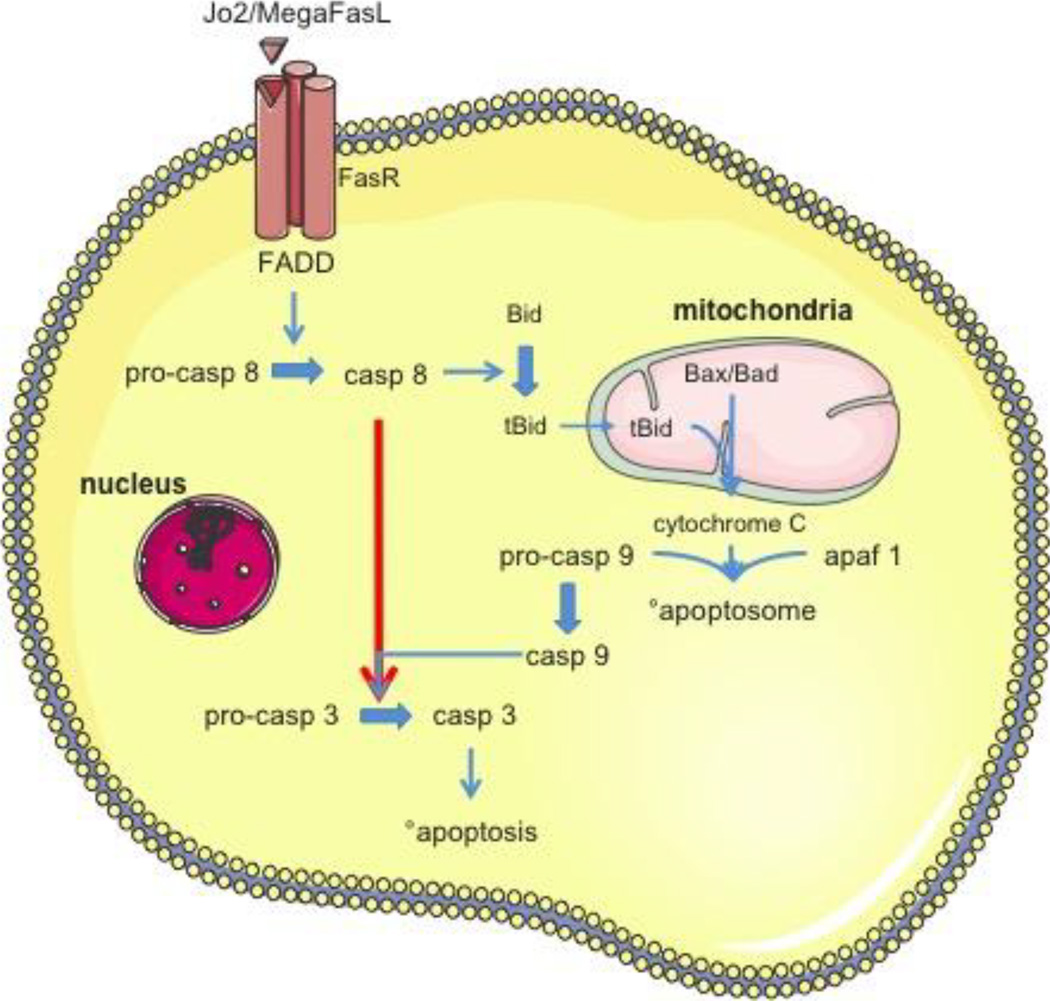
Keywords: Acetaminophen; Acute liver failure; Concanavalin A; D-galactosamine; Fas ligand; Hepatotoxicity.


Conflict of interest statement

Conflict of interest

The authors declare that they have no conflict of interest.

Figures

Figure 1
Figure 1. Chemical structure of acetaminophen/paracetamol
Figure 2
Figure 2. Mechanism of acetaminophen-induced hepatotoxicity
High dose of acetaminophen (APAP) results in the formation of the reactive metabolite N-acetyl-p-benzoquinone imine (NAPQI) by cytochrome P450 (CYP) enzymes in hepatocytes. NAPQI depletes the glutathione (GSH) pool and binds to cysteine sulfhydryl groups of proteins thereby forming APAP-protein adducts. This initiates mitochondrial oxidant stress and peroxynitrite formation. The early oxidant stress triggers activation of mitogen-activated protein kinases (MAPK) that ultimately cause c-jun N-terminal kinase activation (p-JNK), which then translocates to mitochondria to further exacerbate the mitochondrial oxidant stress. The oxidant stress promotes the mitochondrial permeability transition (MPT) pore opening, resulting in the collapse of the membrane potential and cessation of adenosine triphosphate (ATP) synthesis. Early formation of a Bax-based pore in the outer mitochondrial membrane and subsequent swelling of the matrix with rupture of the outer mitochondrial membrane leads to release of intermembrane proteins, such as endonuclease G and apoptosis-inducing factor (AIF), and their translocation to the nucleus with nuclear DNA fragmentation. The massive mitochondrial dysfunction and nuclear DNA damage are the main causes of necrotic cell death.
Figure 3
Figure 3. Chemical structure of D-galactosamine
Figure 4
Figure 4. Pathophysiology of D-galactosamine/endotoxin-based liver injury
Endotoxin (ET) binds to Tolllike receptor 4 (TLR-4) on Kupffer cells, which triggers transcriptional activation of tumor necrosis factor α (TNFα). This results in activation of neutrophils, whereby TNFα is mainly responsible for neutrophil recruitment into the liver sinusoids. Furthermore, TNFα induces various adhesion molecules, such as the intercellular adhesion molecule 1 (ICAM-1), the vascular adhesion molecule 1 (VCAM-1) and selectin chemokines in hepatocytes and endothelial cells. Some of these adhesion molecules are critical for neutrophil extravasation. High dose of D-galactosamine (Gal) depletes the cellular uridine triphosphate and inhibits mRNA synthesis, such as of anti-apoptotic genes, in hepatocytes. This results in activation of the caspase cascade and DNA fragmentation. Caspase activation is also caused by TNFα-induced apoptosis involving the TNFα receptor 1 (TNFα-R1) and caspase activation.
Figure 5
Figure 5. Fas ligand-induced apoptosis in type I and type II cells
Binding of Jo2 or MegaFasL to the Fas ligand receptor (FasR) with a so-called ‘death domain’ (FADD) activates caspase (casp) 8. In type I cells (red arrow), the activation of casp 8 is sufficient to directly activate effector caspase 3. In contrast, in type II cells (blue arrows) casp 8 activation triggers Bid cleavage and truncated Bid (tBid) translocation to mitochondria. Together with Bax and Bad, tBid causes the mitochondrial outer membrane permeabilization with release of cytochrome c. The latter, together with pro-casp 9 and apoptotic protease activating factor 1 (apaf 1), form the apoptosome, which activates casp 9. This leads to drastically enhanced casp 3 activation, followed by induction of apoptosis.

References

    1. Abe K, Ohira H, Kobayashi H, Rai T, Saito H, Takahashi A, Sato Y. Role of CpG ODN in concanavalin A-induced hepatitis in mice. Fukushima J Med Sci. 2005;51:41–49. - PubMed
    1. Abou-Elella AM, Siendones E, Padillo J, Montero JL, De la Mata M, Muntane Relat J. Tumour necrosis factor-alpha and nitric oxide mediate apoptosis by D-galactosamine in a primary culture of rat hepatocytes: exacerbation of cell death by cocultured Kupffer cells. Can J Gastroenterol. 2002;16:791–799. - PubMed
    1. Adamson GM, Harman AW. Oxidative stress in cultured hepatocytes exposed to acetaminophen. Biochem Pharmacol. 1993;45:2289–2294. - PubMed
    1. Albano E, Rundgren M, Harvison PJ, Nelson SD, Moldéus P. Mechanisms of N-acetyl-p-benzoquinone imine cytotoxicity. Mol Pharmacol. 1985;28:306–311. - PubMed
    1. Aninat C, et al. Expression of cytochromes P450, conjugating enzymes and nuclear receptors in human hepatoma HepaRG cells. Drug Metab Dispos. 2006;34:75–83. - PubMed

Publication types