. 2015 Dec 3:5:17622.
doi: 10.1038/srep17622.

TLR-3 stimulation improves anti-tumor immunity elicited by dendritic cell exosome-based vaccines in a murine model of melanoma

Affiliations

TLR-3 stimulation improves anti-tumor immunity elicited by dendritic cell exosome-based vaccines in a murine model of melanoma

Martina Damo et al. Sci Rep. .

Abstract

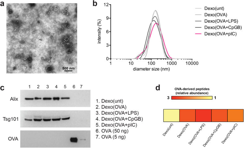
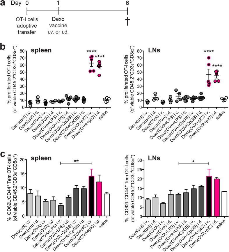
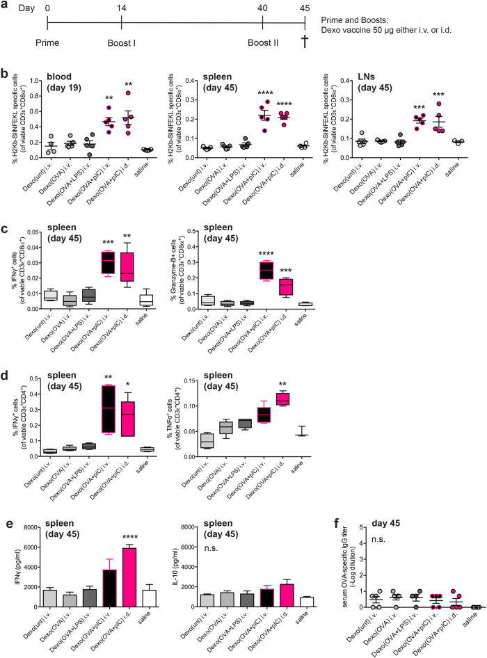
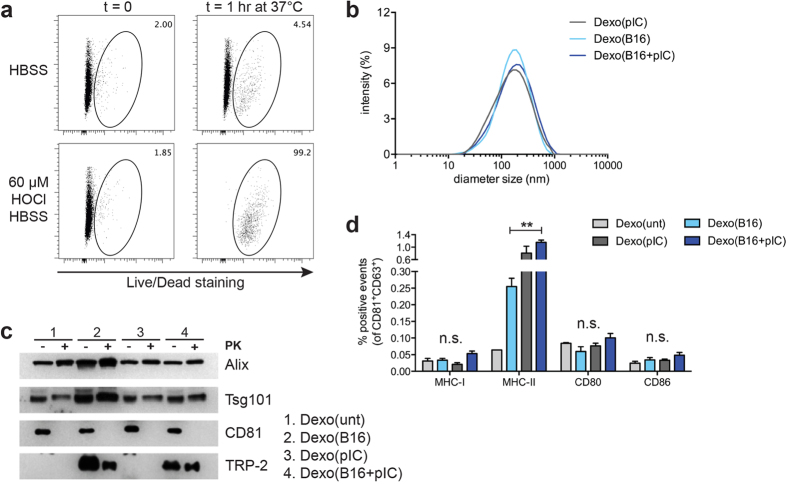
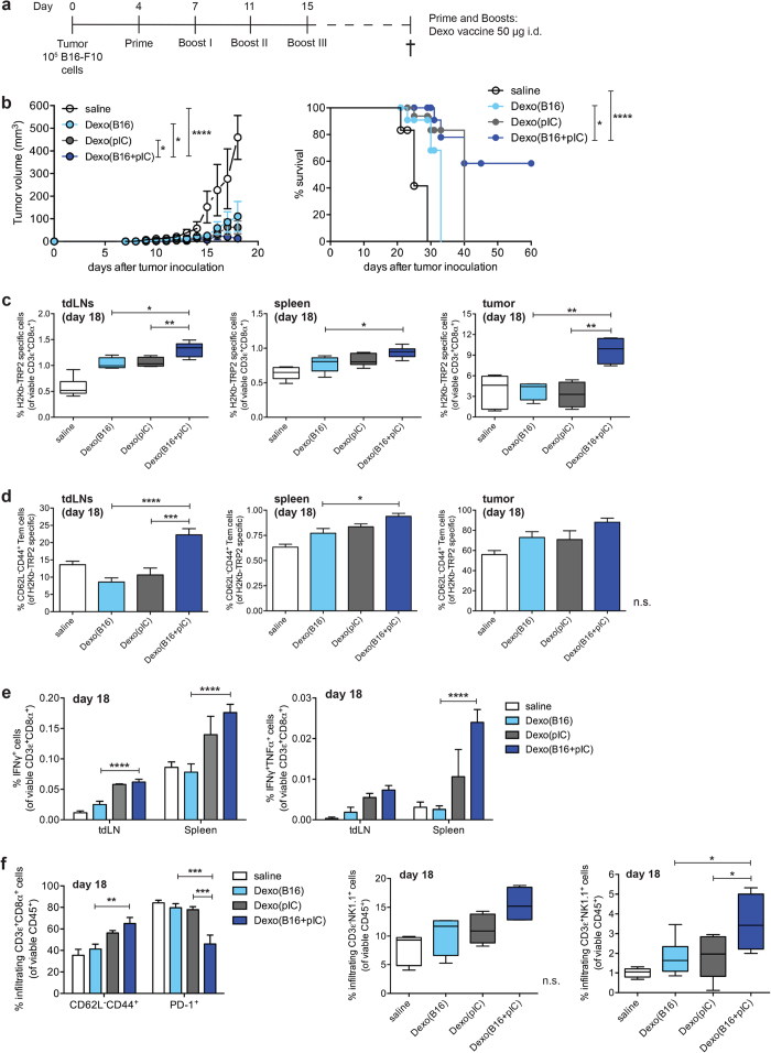
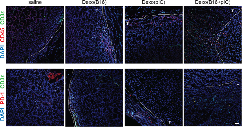
Dendritic cell (DC)-derived exosomes (Dexo) contain the machinery necessary to activate potent antigen-specific immune responses. As promising cell-free immunogens, Dexo have been tested in previous clinical trials for cancer vaccine immunotherapy, yet resulted in limited therapeutic benefit. Here, we explore a novel Dexo vaccine formulation composed of Dexo purified from DCs loaded with antigens and matured with either the TLR-3 ligand poly(I:C), the TLR-4 ligand LPS or the TLR-9 ligand CpG-B. When poly(I:C) was used to produce exosomes together with ovalbumin (OVA), the resulting Dexo vaccine strongly stimulated OVA-specific CD8(+) and CD4(+) T cells to proliferate and acquire effector functions. When a B16F10 melanoma cell lysate was used to load DCs with tumor antigens during exosome production together with poly(I:C), we obtained a Dexo vaccine capable of inducing robust activation of melanoma-specific CD8(+) T cells and the recruitment of cytotoxic CD8(+) T cells, NK and NK-T cells to the tumor site, resulting in significantly reduced tumor growth and enhanced survival as compared to a Dexo vaccine formulation similar to the one previously tested on human patients. Our results indicate that poly(I:C) is a particularly favorable TLR agonist for DC maturation during antigen loading and exosome production for cancer immunotherapy.


Figures

Figure 1
Figure 1. Exosomes are purified from the supernatant of DCs cultured in the presence of the model antigen ovalbumin and activated with different Toll-like receptor ligands.
Exosomes were purified from the supernatant of untreated BMDCs (Dexo(unt)) or of BMDCs treated with OVA (Dexo(OVA)) or from the supernatant of BMDCs treated with OVA and matured with LPS (Dexo(OVA + LPS)), CpG-B (Dexo(OVA + CpGB) or poly(I:C) (Dexo(OVA + pIC)). (a) Example of transmission electron microscopy of one of the Dexo samples after purification from the supernatant of BMDCs confirms the expected physical characteristics of exosomal vesicles. (b) Diameter of exosomes was measured by DLS analysis of the different Dexo samples. (c) Presence of the exosome-specific markers Alix (100 kDa) and Tsg101 (46 kDa) and of full-length OVA protein (45 kDa) was detected by western blot to confirm the identity of bona-fide exosomes and loading of intact OVA in Dexo samples. 50 ng and 5 ng of OVA were used as positive control for detection of full-length OVA. (d) Presence of OVA-derived peptides was detected in Dexo samples by mass spectrometry. Abundance of OVA-related peptides in Dexo(OVA), Dexo(OVA + LPS), Dexo(OVA + CpGB) and Dexo(OVA + pIC) samples is normalized to the background abundance of OVA-related peptides detected in Dexo(unt) samples (relative abundance).
Figure 2
Figure 2. Vaccination with exosomes from OVA-loaded and poly(I:C)-activated DCs strongly activates proliferation and acquisition of effector functions of adoptively transferred OT-I OVA-specific CD8+ T cells in vivo.
(a) CFSE-labeled OT-I CD8+ T cells (CD45.2+) were intravenously transferred into CD45.1+ recipient mice on day 0. Recipient mice were vaccinated on day 1 with 50 μg of the indicated Dexo formulation or saline. On day 6, spleens and LNs were collected to analyze OT-I cells. (b) Proliferation was measured by flow cytometry as dilution of CFSE dye in adoptively transferred CD45.2 + CD3ε +CD8α+ OT-I cells retrieved from the spleen (left) and LNs (right). (c) Acquisition of the CD62L-CD44+ effector memory phenotype of adoptively transferred CD45.2+ CD3ε+ CD8α+ OT-I cells was measured by flow cytometric analysis of cells harvested from the spleen (left) and LNs (right). Data represent mean ± SEM from 2 independent experiments (N = 10). Statistical analysis was performed by one-way ANOVA and Bonferroni post-hoc test correction. In (b) statistics represent comparisons between Dexo(OVA + pIC) i.v. with both Dexo(OVA + LPS) i.v. and Dexo(OVA + CpGB) i.v. groups or between Dexo(OVA + pIC) i.d. with both Dexo(OVA + LPS) i.d. and Dexo(OVA + CpGB) i.d. groups. In (c) statistics represent comparisons between the indicated experimental groups. *P < 0.05 **P < 0.01 and ****P < 0.0001.
Figure 3
Figure 3. Vaccination with exosomes from OVA-loaded and poly(I:C)-activated DCs induces the expansion and acquisition of effector functions of endogenous OVA-specific CD4+ and CD8+ T cells with negligible OVA-specific antibody titers in vivo.
(a) Wild-type mice were vaccinated with 50 μg of the indicated Dexo formulation or saline on day 0 (prime), 14 (boost I) and 40 (boost II). On day 45, spleens and LNs were collected to analyze OVA-specific T cell responses, and blood was sampled to measure the titer of OVA-specific IgG antibodies in the serum. (b) Pentamer staining and flow cytometric analysis were used to measure the frequency of SIINFEKL-specific CD8+ T lymphocytes in the blood of vaccinated mice on day 19 (left) or at the end of the experimental time on day 45 in the spleen (middle) and LNs (right) in the population of viable CD3ε+CD8α+ cells. (c) Splenocytes from vaccinated mice were collected on day 45 to measure acquisition of effector functions by SIINFEKL-specific CD8+ T lymphocytes as detected by intracellular staining for IFNγ (left) and Granzyme-B (right) and flow cytometric analysis. (d) Splenocytes from vaccinated mice were collected on day 45 to measure acquisition of effector functions by OVA-specific CD4+ T lymphocytes as detected by intracellular staining for IFNγ (left) and TNFα (right) and flow cytometric analysis. (e) Splenocytes from vaccinated mice were collected on day 45 to measure IFNγ or IL-10 secreted in the cell supernatant by ELISA upon restimulation in the presence of OVA. (f) Blood from vaccinated mice was collected on day 45 and the titer of OVA-specific IgG antibodies in the serum was measured by ELISA. Data represent mean ± SEM from 2 independent experiments (N = 10). Statistical analysis was performed by one-way ANOVA and Bonferroni post-hoc test correction. *P < 0.05 **P < 0.01, ***P < 0.001, ****P < 0.0001 and n.s. = not significant for comparisons of Dexo(OVA + pIC) administered i.v. or i.d. with Dexo(OVA + LPS).
Figure 4
Figure 4. HOCl-oxidized B16-F10 melanoma cells can be used as a source of tumor antigens for the production of DC exosomes containing melanoma-derived epitopes.
(a) B16-F10 melanoma cells were resuspended in 60 μM HOCl HBSS buffer and incubated at 37 °C for 1 hr to induce oxidation of the tumor cells. After incubation, cells were stained with a viability dye and analyzed by flow cytometry. As controls, viability of B16-F10 cells before incubation (t = 0) or B16-F10 cells resuspended in HBSS buffer was also analyzed. Numbers indicate the frequency of dead cells gated in the total population of B16-F10 cells. (b) Dexo were purified from the supernatant of DCs preincubated with oxidized B16-F10 obtained as in (a) with or without poly(I:C) (Dexo(B16 + pIC) and Dexo(B16), respectively) or with poly(I:C) only as a control (Dexo(pIC)) following the described protocol for exosomes isolation. After purification, the size of Dexo was measured by DLS analysis. (c) Presence of the exosome-specific markers Alix (100 kDa) and Tsg101 (46 kDa) (intravesicular) and CD81 (26 KDa) (vesicle membrane) and of the full-length melanoma-derived protein TRP-2 (59 kDa) was detected by western blot analysis of Dexo(unt), Dexo(B16), Dexo(pIC) and Dexo(B16 + pIC) digested or not with proteinase K to confirm purification of bona-fide exosomes and exosomal localization of the antigens. (d) Surface staining for MHC-I, MHC-II, CD80 and CD86 and flow cytometric analysis of Dexo(unt), Dexo(B16), Dexo(pIC) and Dexo(B16 + pIC). Percentages represent the frequency of positive events among CD81+ CD63+ particles. Data in (d) represent mean ± SEM from 2 independent experiments (N = 6). Statistical analysis was performed by one-way ANOVA and Bonferroni post-hoc test correction to compare Dexo(B16), Dexo(pIC) and Dexo(B16 + pIC). **P < 0.01 and n.s. = not significant.
Figure 5
Figure 5. Therapeutic vaccination with tumor cell antigen-loaded exosomes from poly(I:C)-activated DCs significantly reduces the growth of B16-F10 tumors and improves the survival of tumor-bearing mice by activating melanoma-specific CD8+ T cells and promoting tumor infiltration of cytotoxic cells.
(a) B16-F10 melanoma cells were inoculated subcutaneously between the scapulae of C57BL/6 mice on day 0. To target the tdLNs, recipient mice were vaccinated i.d. in the frontal footpads with 50 μg of the indicated Dexo formulation or saline on day 4 (prime), 7 (boost I), 11 (boost II) and 15 (boost III). (b) Tumor volumes (left) and survival rate (right) were measured from day 0 to day 18 and from day 0 to day 60 after tumor inoculation, respectively. (c) Frequencies of TRP-2180–188 (SVYDFFVWL)-specific CD8+ T lymphocytes in the population of viable CD3ε+CD8α+ cells harvested on day 18 from the tdLNs (left), spleen (middle) and tumor mass (right). (d) Frequencies of CD62L-CD44+ effector memory TRP2-specific CD8+ T lymphocytes harvested on day 18 from the tdLNs (left), spleen (middle) and tumor mass (right). (e) Acquisition of effector functions by B16-F10-specific CD8+ T lymphocytes as indicated by intracellular staining of IFNγ and TNFα and flow cytometric analysis. (f) Frequencies of tumor-infiltrating CD62L-CD44+ effector memory and exhausted PD-1+ CD8+T lymphocytes in the population of total viable CD45+CD3ε+CD8α+ cells (left). Frequencies of tumor-infiltrating NK cells (middle) and NK-T cells (right) among viable CD45+ CD3ε-NK1.1+ or CD45+ CD3ε+ NK1.1+ cells, respectively. Data represent mean ± SEM from 2 independent experiments (N = 15). Statistical analysis of tumor volumes in (b) was performed by unpaired t test comparing Dexo(B16 + pIC) group with either saline, Dexo(B16) or Dexo(pIC) group. *P < 0.1; ****P < 0.0001. Statistical analysis of survival rates in (b) was performed by Log-rank Mantel-Cox test. *P < 0.05; ****P < 0.0001. Statistical analysis of data in (df) was performed by one-way ANOVA and Bonferroni post-hoc test correction. *P < 0.05, **P < 0.01, ***P < 0.001, ****P < 0.0001 and n.s. = not significant for comparisons of Dexo(B16 + pIC) with Dexo(B16) and Dexo(pIC).
Figure 6
Figure 6. Vaccination with tumor cell antigen-loaded exosomes from poly(I:C)-activated DCs induces massive recruitment and infiltration of the tumor mass by PD-1 lymphocytes.
Tumor sections from C57BL/6 mice vaccinated as indicated in Fig. 5a with the indicated Dexo formulation or saline and euthanized on day 18 were stained for either CD45 and CD3ε (top) or CD45 and PD-1 (bottom) and imaged by confocal microscopy. T = intratumoral area. Representative images from 2 independent experiments are shown (N = 15). Scale bar = 50 μm.

References

    1. Gajewski T. F. Cancer immunotherapy. Mol. Oncol. 6, 242–250 (2012). - PMC - PubMed
    1. Mellman I., Coukos G. & Dranoff G. Cancer immunotherapy comes of age. Nature 480, 480–489 (2011). - PMC - PubMed
    1. Mac Keon S., Ruiz M. A. S., Gazzaniga S. & Wainstok R. Dendritic cell-based vaccination in cancer: therapeutic implications emerging from murine models. Front. Immunol. 6, 1–18 (2015). - PMC - PubMed
    1. Palucka K. & Banchereau J. Cancer immunotherapy via dendritic cells. Nat. Rev. Cancer 12, 265–277 (2012). - PMC - PubMed
    1. Chaput N. et al. Dendritic cell derived-exosomes: biology and clinical implementations. J. Leukoc. Biol. 80, 471–478 (2006). - PubMed

Publication types