. 2016 Feb 5;291(6):2954-66.
doi: 10.1074/jbc.M115.683292. Epub 2015 Dec 2.

The Prodomain-bound Form of Bone Morphogenetic Protein 10 Is Biologically Active on Endothelial Cells

Affiliations

The Prodomain-bound Form of Bone Morphogenetic Protein 10 Is Biologically Active on Endothelial Cells

He Jiang et al. J Biol Chem. .

Abstract

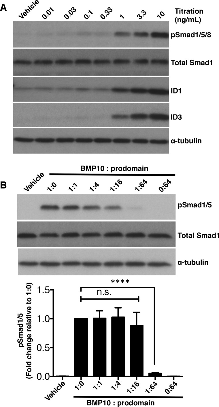
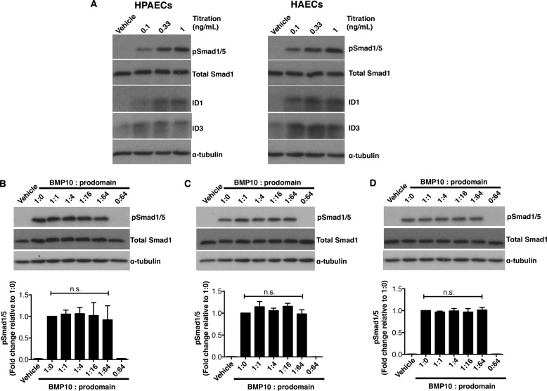
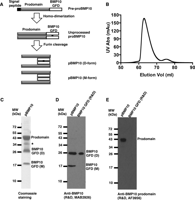
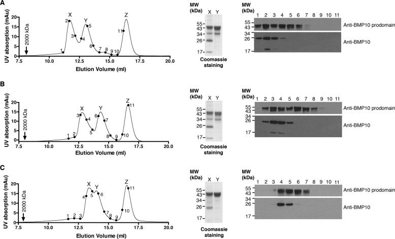
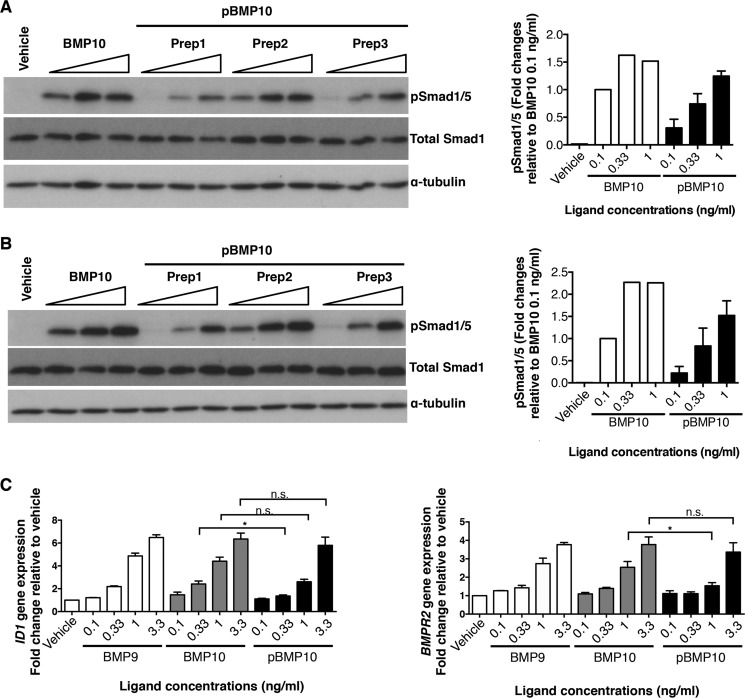
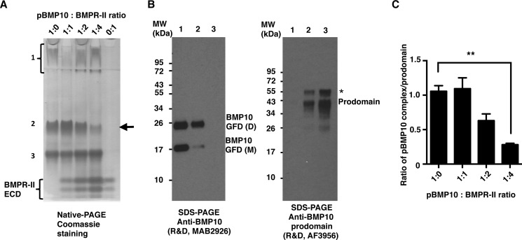
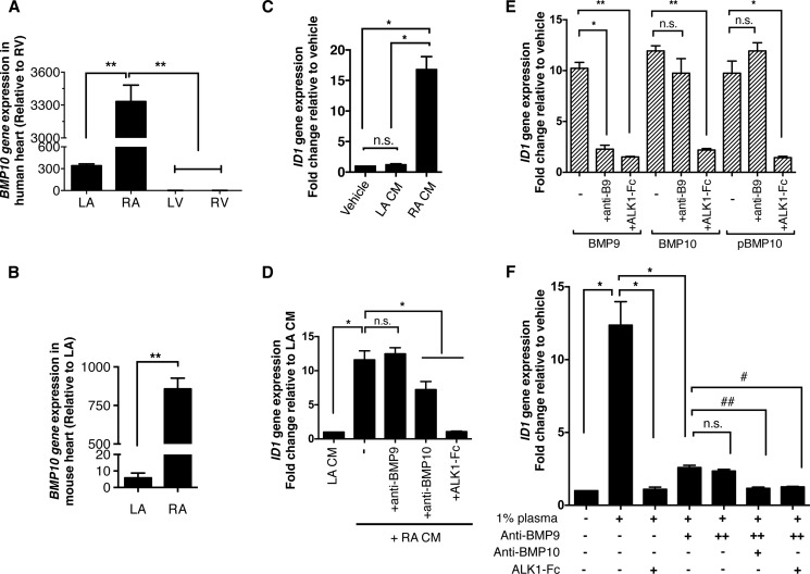
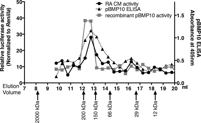
BMP10 is highly expressed in the developing heart and plays essential roles in cardiogenesis. BMP10 deletion in mice results in embryonic lethality because of impaired cardiac development. In adults, BMP10 expression is restricted to the right atrium, though ventricular hypertrophy is accompanied by increased BMP10 expression in a rat hypertension model. However, reports of BMP10 activity in the circulation are inconclusive. In particular, it is not known whether in vivo secreted BMP10 is active or whether additional factors are required to achieve its bioactivity. It has been shown that high-affinity binding of the BMP10 prodomain to the mature ligand inhibits BMP10 signaling activity in C2C12 cells, and it was proposed that prodomain-bound BMP10 (pBMP10) complex is latent. In this study, we demonstrated that the BMP10 prodomain did not inhibit BMP10 signaling activity in multiple endothelial cells, and that recombinant human pBMP10 complex, expressed in mammalian cells and purified under native conditions, was fully active. In addition, both BMP10 in human plasma and BMP10 secreted from the mouse right atrium were fully active. Finally, we confirmed that active BMP10 secreted from mouse right atrium was in the prodomain-bound form. Our data suggest that circulating BMP10 in adults is fully active and that the reported vascular quiescence function of BMP10 in vivo is due to the direct activity of pBMP10 and does not require an additional activation step. Moreover, being an active ligand, recombinant pBMP10 may have therapeutic potential as an endothelial-selective BMP ligand, in conditions characterized by loss of BMP9/10 signaling.

Keywords: bone morphogenetic protein (BMP); bone morphogenetic protein 10 (BMP10); cell biology; endothelial cell; signal transduction; transforming growth factor beta (TGF-β).


Figures

FIGURE 1.
FIGURE 1.
BMP10 prodomain inhibits BMP10 activity in C2C12 cells. A, titration of BMP10 GFD activity in C2C12 cells. Serum-starved C2C12 cells were treated with BMP10 GFD at increasing concentrations for 1 h, and the phosphorylation of Smad1/5 and the induction of ID1 and ID3 proteins were measured by immunoblotting analysis. Total Smad1 was used as a loading control. B, prodomain inhibition assay in C2C12 cells. BMP10 GFD was pre-incubated with the prodomain (molar ratio BMP10 GFD to prodomain, 1:0, 1:1, 1:4, 1:16, and 1:64) before applying to the serum-starved C2C12 cells for 1 h. Ratio 0:64 indicates the same amount of prodomain as in 1:64, but in the absence of BMP10 GFD. The remaining activity was measured by phosphorylation of Smad1/5 using immunoblotting; total Smad1 was used as a loading control. One representative blot from four repeats is shown. Below, using densitometry analysis (Image J), relative phosphorylation of Smad1/5 were corrected to total Smad1 and normalized to the sample treated with BMP10 only without the prodomain (1:0). The fold changes are expressed as mean ± S.E. n = 4; ****, p ≤ 0.0001; n.s., not significant. One-way ANOVA, Dunnett's post test.
FIGURE 2.
FIGURE 2.
BMP10 prodomain does not inhibit BMP10 activity in endothelial cell lines. A, titration of BMP10 GFD activities in HPAECs (left) and HAECs (right). Increasing concentrations of BMP10 GFD as indicated were used to treat the serum-starved cells. After 1 h treatment, cells were harvested and the phosphorylation of Smad1/5 and the induction of ID1 and ID3 proteins were analyzed using immunoblotting. B, BMP10 GFD was pre-incubated with increasing amounts of BMP10 prodomain in same molar ratio as in Fig. 1B before applying to serum-starved endothelial cells in (B) HPAECs, (C) HAECs, and (D) HMEC-1. Remaining activity of BMP10 was measured by phosphorylation of Smad1/5 with total Smad1 as a loading control. One representative blot from four repeats is shown for all experiments. Prodomain inhibitions were quantified and analyzed as in Fig. 1B and shown below.
FIGURE 3.
FIGURE 3.
Generation of recombinant human pBMP10. A, schematic diagram of BMP10 production and processing. B, FPLC chromatography gel filtration trace of purified pBMP10. C, purified pBMP10 shown as the peak fraction from the gel filtration in B on a non-reducing SDS-PAGE. A single asterisk denotes a nonspecific protein. D, both D- and M-forms of BMP10 GFD could be detected by monoclonal anti-BMP10 antibody. E, prodomain can be detected by anti-BMP10 prodomain antibody. BMP10 GFD from R&D Systems was used as a positive control in D and negative control in E.
FIGURE 4.
FIGURE 4.
Prodomain-bound BMP10 is a highly stable complex. Analytical gel filtration analysis of semi-purified pBMP10 in TBS (A), TBS with NaCl at 1 m final concentration (B) or TBS with 1 m GuHCl (C). pBMP10 was pre-incubated in each buffer for 30 min before being loaded onto a Superdex S200 10/300 size-exclusion column pre-equilibrated in the same buffer. A control protein carbonic anhydrase (24 kDa) that does not interact with either BMP10 GFD or its prodomain was added to the pBMP10 before loading. Blue dextran (2000 kDa, black arrow) was run separately in each buffer to indicate the void volume. Note the protein peaks shifted slightly between the runs, potentially due to proteins interacting with the column matrix differently in different buffer systems. Points 1–11 on the traces correspond to consecutive fractions, which were run in lanes 1–11 of immunoblotting analyses probed with either anti-BMP10 prodomain or anti-BMP10 antibodies. Peaks X and Y were TCA precipitated, ran on a non-reducing SDS-PAGE and Coomassie Blue stained to reveal the identity of the peaks. Peak Z is carbonic anhydrase.
FIGURE 5.
FIGURE 5.
Prodomain-bound BMP10 is active in endothelial cells. A and B, phosphorylation of Smad1/5 in HPAECs (A) and HAECs (B) treated with 0.1 ng/ml, 0.33 ng/ml, and 1 ng/ml of BMP10 GFD or three independent preparations of pBMP10 for 1 h, detected by Smad1/5 phosphorylation in immunoblotting analysis; total Smad1 was used as a loading control. The concentrations of pBMP10 in all the cell assays refer to the concentrations of mature GFD in the pBMP10 complex. Relative phosphorylation of Smad1/5 upon treatment was measured using densitometry, corrected to total Smad1 and normalized to 0.1 ng/ml BMP10 GFD treatment condition and plotted on the right. C, induction of ID1 and BMPR2 mRNA expression by recombinant pBMP10, compared with BMP9 and BMP10 GFD. HPAECs were treated with the ligands at indicated concentrations for 8 h before samples were harvested for RNA extraction and qPCR analysis as described in “Experimental Procedures.” n = 3; *, p ≤ 0.05; n.s., not significant; paired t test.
FIGURE 6.
FIGURE 6.
BMPR-II ECD can release BMP10 GFD from the pBMP10 complex. A and B, interaction between BMPR-II ECD and pBMP10 was investigated using native PAGE. Purified pBMP10 complex was run on a 10% native PAGE (A) either alone (1:0), or with increasing amounts of BMPR-II ECD (molar ratio of pBMP10:BMPR-II ECD, 1:1, 1:2, and 1:4). 0:1 refers to BMPR-II alone control. Prodomain-bound BMP10 complex was separated into three bands on the native PAGE. These three bands were excised from the native PAGE, run in parallel on non-reducing SDS-PAGE, and probed with either anti-BMP10 antibody or anti-BMP10 prodomain antibody (B). Band 1, which ran as a smeared band and stayed in the stacking gel, contains only BMP10 GFD, consistent with the PI of BMP10 GFD being 8.67. Band 3 contained only the prodomain (PI = 4.54) and ran fastest on the SDS-PAGE. Band 2 contained both BMP10 GFD and the prodomain, hence is the pBMP10 complex. The arrow in A points to the decrease in the pBMP10 complex upon adding increasing amounts of BMPR-II ECD. A single asterisk in B denotes either a nonspecific protein in the protein prep or a differentially processed BMP10 prodomain. C, changes of pBMP10 complex in A were quantified as the ratio of the pBMP10 complex/prodomain alone to normalize for the loading. n = 4, one-way ANOVA, Dunnett's post test. **, p ≤ 0.01.
FIGURE 7.
FIGURE 7.
BMP10 derived from atrium or plasma is fully active. A, BMP10 mRNA expression in human heart tissues. n = 3. **, p ≤ 0.01; one-way ANOVA, Tukey's post test. B, BMP10 mRNA expression in mouse heart tissues. BMP10 expression is significantly higher in RA than LA in mouse. n = 4, paired t test; **, p ≤ 0.01; C, BMP activity could be detected in the conditioned medium of cultured mouse RA. Conditioned medium from LA or RA was applied to serum-starved HPAECs (both at 5% v/v), and the BMP activity was measured by the induction of ID1 gene expression. No BMP activity can be detected from LA-conditioned medium (LA CM), while significant level of ID1 gene induction activity can be detected in the RA-conditioned medium (RA CM). n = 3, * p ≤ 0.05; n.s., not significant. One-way ANOVA, Tukey's post test; D, identification of BMP activity in RA CM. The ID1-induction activity from RA CM (0.4% v/v) could not be inhibited by anti-BMP9 antibody (at 20 μg/ml), but can be partially inhibited by anti-BMP10 antibody (at 20 μg/ml), and very effectively inhibited by ALK1-Fc (at 2.5 μg/ml). n = 3, *, p ≤ 0.05; n.s., not significant. One-way ANOVA, Dunnett's post test; E, control experiments showed that anti-BMP9 antibody (at 10 μg/ml) could specifically neutralize BMP9 activity very effectively, but not the activity of BMP10 or pBMP10, whereas ALK1-Fc (at 2.5 μg/ml) can inhibit both BMP9 and BMP10 activity very effectively. The concentrations of BMP9, BMP10, and pBMP10 used in this assay were all 1 ng/ml. n = 3, one-way ANOVA for each BMP ligand group, Dunnett's post test, **, p ≤ 0.01; *, p ≤ 0.05; n.s., not significant; F, BMP10 activity can be detected in human plasma. Freshly frozen human plasma was used to treat serum-starved HPAECs (1% v/v final concentration), and BMP activity was measured by ID1 gene induction. All the ID1-induction activity from 1% plasma can be completely inhibited by ALK1-Fc (at 2.5 μg/ml) alone, suggesting that all the ID1-gene induction activity in 1% plasma was due to BMP9 and BMP10. While most of this activity can be inhibited by anti-BMP9 antibody (at 15 μg/ml), the residual ID1-induction activity cannot be inhibited by additional amounts of anti-BMP9 antibody (at 20 μg/ml). It can be only inhibited by either anti-BMP10 antibody (at 15 μg/ml) or ALK1-Fc (at 2.5 μg/ml), suggesting the residual ID1-induction activity is due to BMP10. n = 3, one-way ANOVA, Dunnett's post test. *, p ≤ 0.05; #, p ≤ 0.05; ##, p ≤ 0.01; n.s., not significant.
FIGURE 8.
FIGURE 8.
Right-atrium-secreted active BMP10 is in the prodomain-bound form. Proteins from RA-conditioned medium were separated by gel filtration chromatography, and the BMP activities in the fractions were measured using the BRE-luciferase assay as described in “Experimental Procedures.” The relative BRE-luciferase activities in the fractions were plotted against their elution volumes (black line with circle symbols). The same process was repeated with diluted, purified recombinant human pBMP10, and the relative BMP activities in the fractions were also measured and plotted (gray line with square symbols). In addition, a pBMP10-specific ELISA was carried out to measure the proteins in the fractions from RA-conditioned medium gel filtration. Data were presented as optical densities (absorbance at 405 nm, right axis, dotted line with triangular symbols). Proteins from Gel Filtration Calibration Kit were run under identical conditions, and arrows below showed the elution volumes of the protein standards.

References

    1. Neuhaus H., Rosen V., and Thies R. S. (1999) Heart specific expression of mouse BMP-10 a novel member of the TGF-β superfamily. Mech. Dev. 80, 181–184 - PubMed
    1. Chen H., Shi S., Acosta L., Li W., Lu J., Bao S., Chen Z., Yang Z., Schneider M. D., Chien K. R., Conway S. J., Yoder M. C., Haneline L. S., Franco D., and Shou W. (2004) BMP10 is essential for maintaining cardiac growth during murine cardiogenesis. Development 131, 2219–2231 - PMC - PubMed
    1. Chen H., Yong W., Ren S., Shen W., He Y., Cox K. A., Zhu W., Li W., Soonpaa M., Payne R. M., Franco D., Field L. J., Rosen V., Wang Y., and Shou W. (2006) Overexpression of bone morphogenetic protein 10 in myocardium disrupts cardiac postnatal hypertrophic growth. J. Biol. Chem. 281, 27481–27491 - PMC - PubMed
    1. Nakano N., Hori H., Abe M., Shibata H., Arimura T., Sasaoka T., Sawabe M., Chida K., Arai T., Nakahara K., Kubo T., Sugimoto K., Katsuya T., Ogihara T., Doi Y., Izumi T., and Kimura A. (2007) Interaction of BMP10 with Tcap may modulate the course of hypertensive cardiac hypertrophy. Am. J. Physiol. Heart Circ. Physiol. 293, H3396–H3403 - PubMed
    1. Mazerbourg S., Sangkuhl K., Luo C. W., Sudo S., Klein C., and Hsueh A. J. (2005) Identification of receptors and signaling pathways for orphan bone morphogenetic protein/growth differentiation factor ligands based on genomic analyses. J. Biol. Chem. 280, 32122–32132 - PubMed

Publication types