. 2016 Jan 15;291(3):1492-500.
doi: 10.1074/jbc.M115.694018. Epub 2015 Dec 2.

DndEi Exhibits Helicase Activity Essential for DNA Phosphorothioate Modification and ATPase Activity Strongly Stimulated by DNA Substrate with a GAAC/GTTC Motif

Affiliations

DndEi Exhibits Helicase Activity Essential for DNA Phosphorothioate Modification and ATPase Activity Strongly Stimulated by DNA Substrate with a GAAC/GTTC Motif

Tao Zheng et al. J Biol Chem. .

Abstract

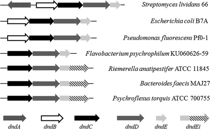
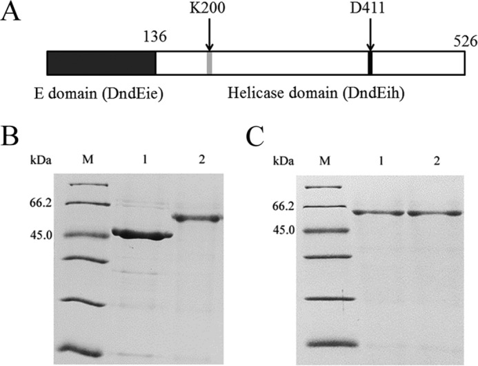
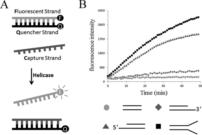
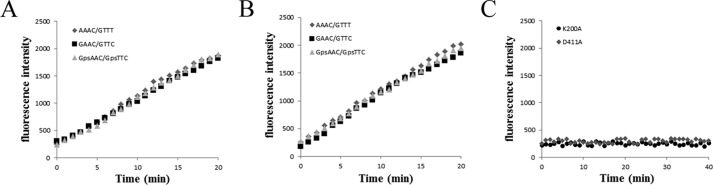
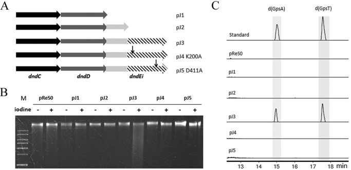
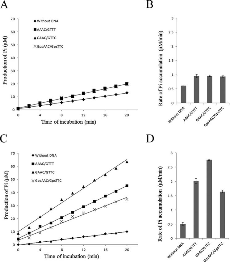
Phosphorothioate (PT) modification of DNA, in which the non-bridging oxygen of the backbone phosphate group is replaced by sulfur, is governed by the DndA-E proteins in prokaryotes. To better understand the biochemical mechanism of PT modification, functional analysis of the recently found PT-modifying enzyme DndEi, which has an additional domain compared with canonical DndE, from Riemerella anatipestifer is performed in this study. The additional domain is identified as a DNA helicase, and functional deletion of this domain in vivo leads to PT modification deficiency, indicating an essential role of helicase activity in PT modification. Subsequent analysis reveals that the additional domain has an ATPase activity. Intriguingly, the ATPase activity is strongly stimulated by DNA substrate containing a GAAC/GTTC motif (i.e. the motif at which PT modifications occur in R. anatipestifer) when the additional domain and the other domain (homologous to canonical DndE) are co-expressed as a full-length DndEi. These results reveal that PT modification is a biochemical process with DNA strand separation and intense ATP hydrolysis.

Keywords: ATPase; DNA enzyme; DNA helicase; bacteria; microbiology.


Figures

FIGURE 1.
FIGURE 1.
Genomic organization of the PT-modifying genes in 7 members of bacteria. Strains and GenBankTM accession numbers: S. lividans 66 (CM001889.1); E. coli B7A (CP005998.1); Pseudomonas fluorescens Pf0–1 (CP000094.2); Flavobacterium psychrophilum KU060626-59 (KF241852); R. anatipestifer ATCC 11845 (CP003388.1); Bacteroides faecis MAJ27 (NZ_AGDG01000022.1); Psychroflexus torquis ATCC 700755 (CP003879.1).
FIGURE 2.
FIGURE 2.
Analysis of the PT modification in R. anatipestifer ATCC 11845. A, electrophoresis pattern of the iodine-cleaved genomic DNA from R. anatipestifer ATCC 11845 (lanes 4 and 5), with R. anatipestifer HXb2 (lacking PT-modifying genes; lanes 2 and 3) as a negative control. M, DNA ladder. B, LC-MS analysis of the PT-linked dinucleotides from R. anatipestifer ATCC 11845.
FIGURE 3.
FIGURE 3.
Purification of DndEih, DndEi, and DndEi mutants. A, domain organization of the DndEi protein. The positions of the two conserved motifs are indicated by gray and black boxes, respectively. The positions of the two point mutations are indicated by arrows. B, SDS-PAGE analysis of purified DndEih and DndEi stained by Coomassie Blue. Lanes: M, molecular mass marker; 1, DndEih; 2, DndEi. C, SDS-PAGE analysis of purified DndEi mutants stained by Coomassie Blue. Lanes: M, molecular mass marker; 1, DndEi (K200A); 2, DndEi (D411A).
FIGURE 4.
FIGURE 4.
DNA helicase activity of DndEih using various DNA substrates. A, schematic representation of the fluorescence helicase assay based on FRET. The fluorescent strand is labeled with a fluorophore (F) and quencher strand is labeled with a quencher (Q). Annealing of the fluorophore strand to the quencher strand results in the fluorescence signal from the Alexa Fluor 488 dye to be quenched. When the double-stranded DNA substrate is unwound by the helicase, the fluorophore emits light upon its release from the quencher. The capture strand, which is complementary to the quencher strand, prevents the reannealing of the unwound duplex. B, time course of the helicase reaction of DndEih using 3′-overhang (diamonds), 5′-overhang (triangles), blunt-ended (circles), and forked (squares) DNA substrates in the presence of ATP.
FIGURE 5.
FIGURE 5.
Single-stranded DNA translocation activity of DndEih. Translocation activity was investigated by measuring the ability of DndEih to displace streptavidin from a biotinylated probe at the 5′ or 3′ end of ssDNA. The black circle indicates the biotin-labeled DNA end, with the larger circle representing the streptavidin. Lane 1, biotinylated DNA; lane 2, biotinylated DNA bound to streptavidin; lane 3, streptavidin-bound DNA incubated with DndEih for 30 min in the absence of ATP; lane 4, streptavidin-bound DNA incubated with DndEih for 30 min in the presence of ATP.
FIGURE 6.
FIGURE 6.
DNA helicase activity of DndEih, DndEi, and DndEi mutants. A and B, time course of the helicase reaction of DndEih (A) and DndEi (B) using 5′-overhang DNA substrates with different sequence motif as indicated. C, time course of the helicase reaction of DndEi mutants using 5′-overhang DNA substrates.
FIGURE 7.
FIGURE 7.
Analysis of the PT modification in R. anatipestifer HXb2 harboring a pReS0-based plasmid (pJ1–5), which has the PT-modifying genes from R. anatipestifer ATCC 11845. A, schematic diagram of the PT-modifying genes in pReS0-based plasmids (Table 1). B, electrophoresis pattern of the iodine-cleaved genomic DNA from R. anatipestifer HXb2 (pJ1) (lanes 4 and 5), HXb2 (pJ2) (lanes 6 and 7), HXb2 (pJ3) (lanes 8 and 9), HXb2 (pJ4) (lanes 10 and 11), HXb2 (pJ5) (lanes 12 and 13), and HXb2 (pReS0 empty vector) (lanes 2 and 3). M, DNA ladder. C, LC-MS analysis of the PT-linked dinucleotides from R. anatipestifer HXb2 harboring a pReS0-based plasmid. Electrospray ionization: m/z [M + H]+ modes for LC-MS: d(GpsA), 597.1388; d(GpsT), 588.1272.
FIGURE 8.
FIGURE 8.
ATPase activity of DndEih (A and B) and DndEi (C and D). A and C, representative time courses of ATP hydrolysis by DndEih and DndEi. The ATPase assay was performed in the absence of DNA or in the presence of different kinds of dsDNA substrate (with no GAAC/GTTC motif; with a GAAC/GTTC motif; with a PT-modified GpsAAC/GpsTTC motif). The amount of Pi produced was calculated based on a standard KH2PO4 curve. Each data point represents the mean of triplicate experiments. B, rates of Pi accumulation in the ATPase assay performed in A. The data are presented as mean ± S.D. D, rates of Pi accumulation in the ATPase assay performed in C. The data are presented as mean ± S.D.
FIGURE 9.
FIGURE 9.
Phylogenetic analysis of DndE homologues, using MEGA v5.0 with 500 bootstrap replicates. Ampersand: DndE; asterisk: the E domain of DndEi (DndEie). GenBankTM accession numbers of the DndE homologues are: C. canimorsus (WP_041913577.1); R. anatipestifer ATCC 11845 (ADQ81723.1); Vibrio cholerae BJG-01 (EGS71244.1); Gelidibacter mesophilus (WP_027127205); Methylobacterium extorquens (WP_012754233); Klebsiella oxytoca (WP_046878204.1); C. perfringens (WP_003471805); Desulfovibrio africanus (PCS EMG37157); Methanosarcina mazei (KKI02757.1); S. cyanosphaera (WP_041620172.1); P. histicola (WP_036868676.1); Flexibacter litoralis (WP_041264540); and Anabaena cylindrica PCC 7122 (AFZ60204.1).

References

    1. Wang L., Chen S., Xu T., Taghizadeh K., Wishnok J. S., Zhou X., You D., Deng Z., and Dedon P. C. (2007) Phosphorothioation of DNA in bacteria by dnd genes. Nat. Chem. Biol. 3, 709–710 - PubMed
    1. Wang L., Chen S., Vergin K. L., Giovannoni S. J., Chan S. W., DeMott M. S., Taghizadeh K., Cordero O. X., Cutler M., Timberlake S., Alm E. J., Polz M. F., Pinhassi J., Deng Z., and Dedon P. C. (2011) DNA phosphorothioation is widespread and quantized in bacterial genomes. Proc. Natl. Acad. Sci. U.S.A. 108, 2963–2968 - PMC - PubMed
    1. Xu T., Yao F., Zhou X., Deng Z., and You D. (2010) A novel host-specific restriction system associated with DNA backbone S-modification in Salmonella. Nucleic Acids Res. 38, 7133–7141 - PMC - PubMed
    1. Cao B., Cheng Q., Gu C., Yao F., DeMott M. S., Zheng X., Deng Z., Dedon P. C., and You D. (2014) Pathological phenotypes and in vivo DNA cleavage by unrestrained activity of a phosphorothioate-based restriction system in Salmonella. Mol. Microbiol. 93, 776–785 - PMC - PubMed
    1. Cheng Q., Cao B., Yao F., Li J., Deng Z., and You D. (2015) Regulation of DNA phosphorothioate modifications by the transcriptional regulator DptB in Salmonella. Mol. Microbiol. 97, 1186–1194 - PubMed

Publication types

MeSH terms