. 2016 Jan 22;291(4):1692-1702.
doi: 10.1074/jbc.M115.674408. Epub 2015 Dec 2.

Novel Structural Components Contribute to the High Thermal Stability of Acyl Carrier Protein from Enterococcus faecalis

Affiliations

Novel Structural Components Contribute to the High Thermal Stability of Acyl Carrier Protein from Enterococcus faecalis

Young-Guen Park et al. J Biol Chem. .

Abstract

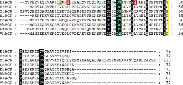
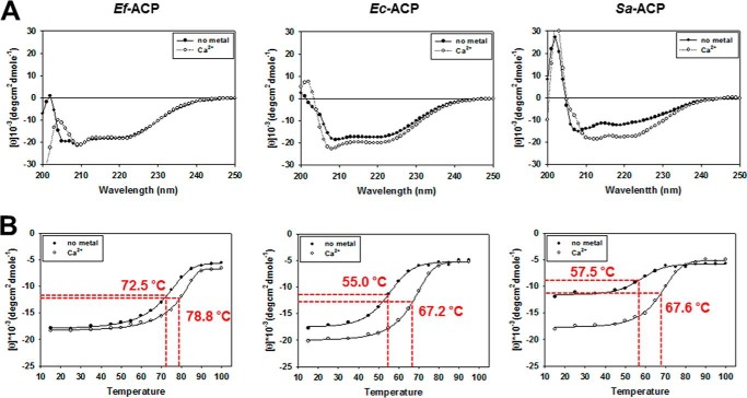
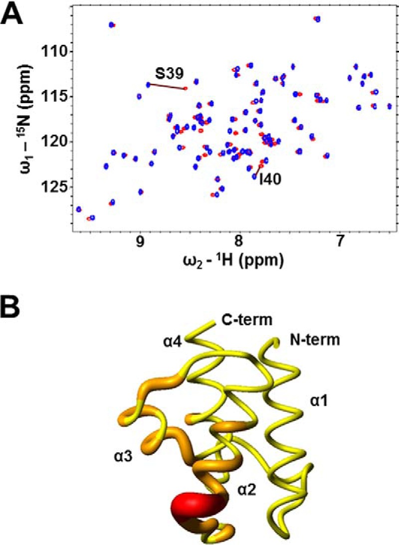
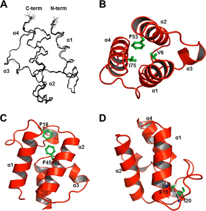
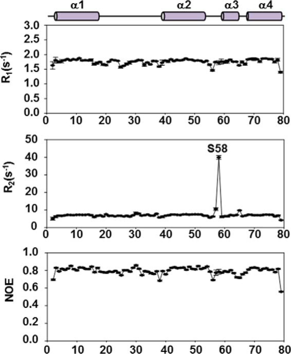
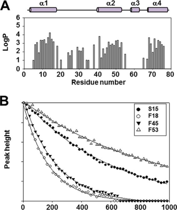
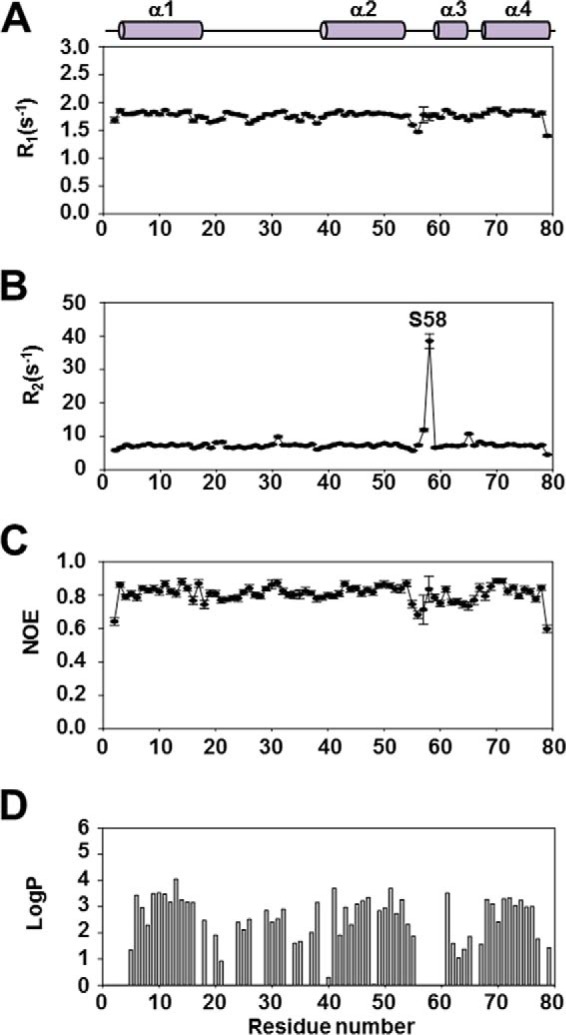
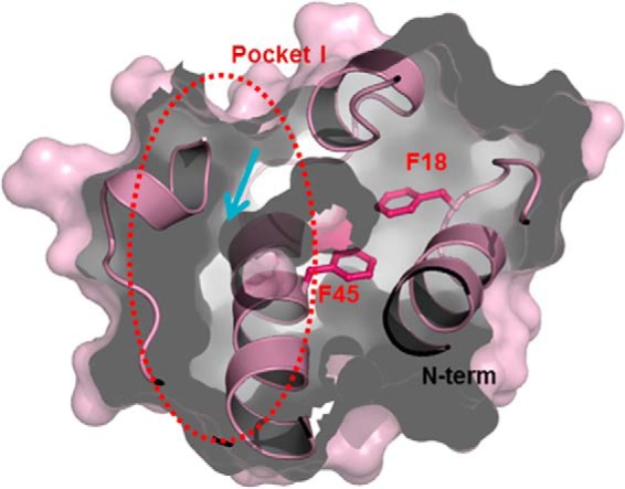
Enterococcus faecalis is a Gram-positive, commensal bacterium that lives in the gastrointestinal tracts of humans and other mammals. It causes severe infections because of high antibiotic resistance. E. faecalis can endure extremes of temperature and pH. Acyl carrier protein (ACP) is a key element in the biosynthesis of fatty acids responsible for acyl group shuttling and delivery. In this study, to understand the origin of high thermal stabilities of E. faecalis ACP (Ef-ACP), its solution structure was investigated for the first time. CD experiments showed that the melting temperature of Ef-ACP is 78.8 °C, which is much higher than that of Escherichia coli ACP (67.2 °C). The overall structure of Ef-ACP shows the common ACP folding pattern consisting of four α-helices (helix I (residues 3-17), helix II (residues 39-53), helix III (residues 60-64), and helix IV (residues 68-78)) connected by three loops. Unique Ef-ACP structural features include a hydrophobic interaction between Phe(45) in helix II and Phe(18) in the α1α2 loop and a hydrogen bonding between Ser(15) in helix I and Ile(20) in the α1α2 loop, resulting in its high thermal stability. Phe(45)-mediated hydrophobic packing may block acyl chain binding subpocket II entry. Furthermore, Ser(58) in the α2α3 loop in Ef-ACP, which usually constitutes a proline in other ACPs, exhibited slow conformational exchanges, resulting in the movement of the helix III outside the structure to accommodate a longer acyl chain in the acyl binding cavity. These results might provide insights into the development of antibiotics against pathogenic drug-resistant E. faecalis strains.

Keywords: Enterococcus faecalis; acyl carrier protein (ACP); biophysics; fatty acid synthase (FAS); nuclear magnetic resonance (NMR); protein structure.


Figures

FIGURE 1.
FIGURE 1.
Sequence alignment of ACP. Sequence alignment of the Ef-ACP protein with three Gram-positive homologues (B. subtilis ACP (BsACP), M. tuberculosis ACP (MtACP), and S. aureus-ACP (SaACP)) and four Gram-negative bacterial homologues (E. coli ACP (EcACP), A. baumanni ACP (AbACP), P. falciparum ACP (PfACP), and V. harveyi ACP (VhACP)). The conserved residues with 100% identity are shown in black. The prosthetic group attachment site, which is highly conserved (Ser39 in Ef-ACP) is marked by a green box. Unique features of Ef-ACP are marked with orange (Ser15), red (Phe18 and Phe45), and yellow boxes (Ser58). The residues marked with a red box form hydrophobic interactions with each other and are found only in Ef-ACP. The residues in the yellow box are composed of a proline in the α2α3 loop of most ACPs, but Ef-ACP carries a serine residue in the α2α3 loop.
FIGURE 2.
FIGURE 2.
CD spectra of Ef-ACP, Ec-ACP, and S. aureus ACP (Sa-ACP). A, CD spectrum of ACPs in the presence of Ca2+ (○) and in the absence of Ca2+ (●) at 25 °C. B, temperature-induced folding change of ACPs as monitored by changes of ellipticity at 222 nm. The red dotted line indicates the Tm point of each graph.
FIGURE 3.
FIGURE 3.
NMR data of apo- and holo-ACP. A, overlay of the 1H-15N HSQC spectra of apo- (red) and holo-ACP (blue). B, chemical shift change value ranges induced by 4′-phosphopantetheine prosthetic group modification of apo-ACP are indicated in various colors in tubular drawings. Residues with ppm >0.03 are shown with graduated larger ribbon diameters. N-term and C-term, N and C termini, respectively. Red, >0.1 ppm; orange, 0.03 < ppm < 0.1; yellow, <0.03 ppm. Chemical shift perturbations were calculated using the equation, Δδav = (0.5(Δδ(1HN)2 + (0.2Δδ(15N))2))½.
FIGURE 4.
FIGURE 4.
Structure of Ef-ACP. A, superposition of backbone (nitrogen, Cα, and C′) atoms of the 20 lowest energy structures of holo-Ef-ACP. All residues (positions 1–79) are shown, and N and C termini are labeled as N-term and C-term, respectively. B, hydrophobic contacts of Val6, Phe53, and Ile75 in holo-Ef-ACP. C, hydrophobic interactions between Phe18 and Phe45. D, hydrogen bond interactions between Ser15 and Ile20 are depicted with black dotted lines. In B–D, all structures are represented as schematic diagrams, and each helix is labeled from α1 to α4. Each residue is depicted as green sticks.
FIGURE 5.
FIGURE 5.
Spin relaxation rates of Ef-ACP in the presence of Ca2+. Relaxation data of Ef-ACP were measured at 298 K. The values of R1, R2, and the steady-state heteronuclear NOEs are plotted as a function of the residue sequence number for Ef-ACP. Above the plots, the solid and filled bars represent the four helices of Ef-ACP.
FIGURE 6.
FIGURE 6.
Hydrogen/deuterium exchange experiment of Ef-ACP in the presence of Ca2+. A, the protection factor of Ef-ACP is depicted by a bar graph as a function of the residue sequence number of Ef-ACP. The solid and filled bars above the plots represent the four helices of Ef-ACP. B, time course of exchange showing signal loss due to hydrogen/deuterium exchange as a function of time after the addition of D2O for each of the four residues. Peak height is in arbitrary units.
FIGURE 7.
FIGURE 7.
Spin relaxation rates and Protection factor of Ef-ACP in the absence of Ca2+. The values of R1 (A), R2 (B), and the steady-state heteronuclear NOEs (C) are plotted as a function of the residue sequence number for Ef-ACP. Above the plots, the solid and filled bars represent the four helices of Ef-ACP. D, protection factor of Ef-ACP in the absence of Ca2+ depicted by a bar graph as a function of the residue sequence number of Ef-ACP. The solid and filled bars above the plots represent the four helices of Ef-ACP. All NMR data of Ef-ACP were measured at 298 K.
FIGURE 8.
FIGURE 8.
Surface representation of subpockets in the hydrophobic cavity. Phe45 in Ef-ACP has hydrophobic interactions with Phe18, resulting in blockage of the entrance into the alternate binding cavity of Ef-ACP between helices I, II, and IV. The arrows represent the direction of the path of the growing acyl chain.
FIGURE 9.
FIGURE 9.
Electrostatic potential surface structure and structural comparison of ACPs. A–C, electrostatic potential of Ec-ACP (PDB entry 2K93) (A), V. harveyi ACP (PDB entry 2L0Q) (B), and Ef-ACP (PDB entry 2N50) (C). The residue numbers are labeled. Positive and negative charge residues are colored in blue and red, respectively. The His17 residue is colored in green. This surface structure is rotated about the y axis by 90° compared with that shown in Fig. 4A. D–G, structural comparison of Ef-ACP with other ACPs; overlay of structures of Ef-ACP (red; PDB entry 2N50) and Ec-ACP (yellow; PDB entry 2K93) (D), Ef-ACP and M. tuberculosis ACP (blue; PDB entry 1KIP) (E), Ef-ACP and P. falciparum ACP (cyan; PDB entry 2QF0) (F), and Ef-ACP and B. subtilis ACP (green; PDB entry 1HY8) (G). Each helix is labeled from α1 to α4, and this overlay structure has the same orientation as the structure shown in Fig. 4A.

References

    1. Uauy R., Mena P., and Rojas C. (2000) Essential fatty acids in early life: structural and functional role. Proc. Nutr. Soc. 59, 3–15 - PubMed
    1. Cronan J. E., Jr. (2004) The structure of mammalian fatty acid synthase turned back to front. Chem. Biol. 11, 1601–1602 - PubMed
    1. Cronan J. E. (2006) Remarkable structural variation within fatty acid megasynthases. Nat. Chem. Biol. 2, 232–234 - PubMed
    1. Chan D. I., and Vogel H. J. (2010) Current understanding of fatty acid biosynthesis and the acyl carrier protein. Biochem. J. 430, 1–19 - PubMed
    1. White S. W., Zheng J., Zhang Y. M., and Rock (2005) The structural biology of type II fatty acid biosynthesis. Annu. Rev. Biochem. 74, 791–831 - PubMed

Publication types

MeSH terms

Substances