Review
. 2016 Jan;18(1):13-24.
doi: 10.1016/j.jcyt.2015.10.008. Epub 2015 Nov 26.

The current landscape of the mesenchymal stromal cell secretome: A new paradigm for cell-free regeneration

Affiliations
Review

The current landscape of the mesenchymal stromal cell secretome: A new paradigm for cell-free regeneration

Vijay Bhaskar Reddy Konala et al. Cytotherapy. 2016 Jan.

Abstract

The unique properties of mesenchymal stromal/stem cells (MSCs) to self-renew and their multipotentiality have rendered them attractive to researchers and clinicians. In addition to the differentiation potential, the broad repertoire of secreted trophic factors (cytokines) exhibiting diverse functions such as immunomodulation, anti-inflammatory activity, angiogenesis and anti-apoptotic, commonly referred to as the MSC secretome, has gained immense attention in the past few years. There is enough evidence to show that the one important pathway by which MSCs participate in tissue repair and regeneration is through its secretome. Concurrently, a large body of MSC research has focused on characterization of the MSC secretome; this includes both soluble factors and factors released in extracellular vesicles, for example, exosomes and microvesicles. This review provides an overview of our current understanding of the MSC secretome with respect to their potential clinical applications.

Keywords: cell therapy; exosomes; mesenchymal stromal cells; regeneration; secretome.


Conflict of interest statement

Disclosure of interests: The authors have no commercial, proprietary, or financial interest in the products or companies described in this article.

Figures

Figure 1
Figure 1
MSCs can be isolated from various tissues including bone marrow, umbilical cord, muscle and tooth root. After in vitro expansion, MSCs can be authenticated as per the guidelines laid down by International Society of Cell Therapy (ISCT). Morphologically, MSCs are fibroblast-like, grow as adherent cultures and are capable of forming colonies; they express a panel of markers: positive for Sca-1, CD105, CD73, CD29 and CD90 and negative for CD31, CD34, CD45 and CD11b. In addition, MSCs have the potential to differentiate into adipocytes, chondrocytes, osteoblasts and other cell types.
Figure 2
Figure 2
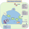
Schematic representation depicting the origin of MVs, exosomes and apoptotic bodies. An MV arises from budding of the plasma membrane. MVs are more irregular in shape and size and can contain cytoplasmic materials; they express surface markers such as integrin-β, CD40 and selectins such as plasma selectins and/or proteins similar to the cells from which they originate. Exosomes are derived from the endosomal trafficking system and therefore are more regular in shape and size. Exosomes are more easily identifiable via cell surface markers such as CD81, CD9 and CD63 and may contain materials such as mitochondrial DNAs, mRNAs and miRNAs. ER, endoplasmic reticulum; miRNA, microRNA; TGN, trans-Golgi network; MVB, multi-vesicular bodies. Apoptotic bodies or vesicles are 1 to 5 μm (>1000 nm) in diameter; they are released from the plasma membrane as blebs when cells undergo apoptosis and contain several intracellular fragments and cellular organelles.
Figure 3
Figure 3
Role of various paracrine factors released by MSCs. Secreted factors may exert different functions on cells via the release of different kinds of molecules, depending on the microenvironment: angiopoietin (Ang), basic fibroblast growth factor (bFGF), brain-derived neurotrophic factor (BDNF), chemokine ligand (CCL), chemokine (C-XC motif) ligand (CXCL), erythropoietin (EPO), glial cell line–neurotrophic factor (GDNF), granulocyte-macrophage–colony-stimulating factor (GM-CSF), hemeoxygenase (HO), hepatocyte growth factor (HGF), human leucocyte antigen (HLA), indoleamine 2,3-dioxygenase (IDO), insulin growth factor (IGF), interleukin (IL), keratinocyte growth factor (KGF), leukemia inhibitory factor (LIF), human cathelicidin (LL37), monocyte chemoattractant protein (MCP), metalloproteinase (MMP), nerve growth factor (NGF), nitric oxide (NO), platelet-derived growth factor (PDGF), prostaglandin (PGE), placental growth factor (PIGF), stem cell factor (SCF), stromal cell–derived factor (SDF), tissue inhibitor of metalloproteinase (TIMP), transforming growth factor (TGF), thrombopoietin (TPO), TNF-α–stimulated gene/protein (TSG) and vascular endothelial growth factor (VEGF).

References

    1. Pittenger MF, Mackay AM, Beck SC, Jaiswal RK, Douglas R, Mosca JD, et al. Multilineage potential of adult human mesenchymal stem cells. Science. 1999;284:143–7. - PubMed
    1. Uccelli A, Moretta L, Pistoia V. Mesenchymal stem cells in health and disease. Nat Rev Immunol. 2008;8(9):726–62. - PubMed
    1. Shi Y, Hu G, Su J, Li W, Chen Q, Shou P, et al. Mesenchymal stem cells: a new strategy for immunosuppression and tissue repair. Cell Res. 2010;20(5):510–18. - PubMed
    1. Davey GC, Patil SB, O’Loughlin A, O’Brien T. Mesenchymal stem cell-based treatment for microvascular and secondary complications of Diabetes mellitus. Front Endocrinol (Lausanne) 2014;5:86. - PMC - PubMed
    1. Nauta AJ, Fibbe WE. Immunomodulatory properties of mesenchymal stromal cells. Blood. 2007;110(10):3499–506. - PubMed

Publication types