

Air-kerma strength determination of a new directional (103)Pd source
- PMID: 26632069
- PMCID: PMC5148138
- DOI: 10.1118/1.4935409
Air-kerma strength determination of a new directional (103)Pd source
Abstract
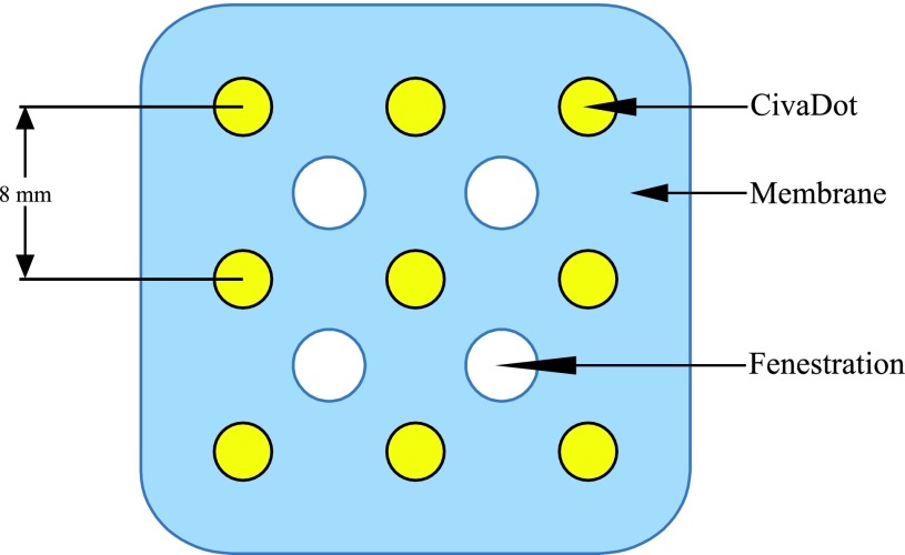
Purpose: A new directional (103)Pd planar source array called a CivaSheet™ has been developed by CivaTech Oncology, Inc., for potential use in low-dose-rate (LDR) brachytherapy treatments. The array consists of multiple individual polymer capsules called CivaDots, containing (103)Pd and a gold shield that attenuates the radiation on one side, thus defining a hot and cold side. This novel source requires new methods to establish a source strength metric. The presence of gold material in such close proximity to the active (103)Pd region causes the source spectrum to be significantly different than the energy spectra of seeds normally used in LDR brachytherapy treatments. In this investigation, the authors perform air-kerma strength (S(K)) measurements, develop new correction factors for these measurements based on an experimentally verified energy spectrum, and test the robustness of transferring S(K) to a well-type ionization chamber.
Methods: S(K) measurements were performed with the variable-aperture free-air chamber (VAFAC) at the University of Wisconsin Medical Radiation Research Center. Subsequent measurements were then performed in a well-type ionization chamber. To realize the quantity S(K) from a directional source with gold material present, new methods and correction factors were considered. Updated correction factors were calculated using the MCNP 6 Monte Carlo code in order to determine S(K) with the presence of gold fluorescent energy lines. In addition to S(K) measurements, a low-energy high-purity germanium (HPGe) detector was used to experimentally verify the calculated spectrum, a sodium iodide (NaI) scintillating counter was used to verify the azimuthal and polar anisotropy, and a well-type ionization chamber was used to test the feasibility of disseminating S(K) values for a directional source within a cylindrically symmetric measurement volume.
Results: The UW VAFAC was successfully used to measure the S(K) of four CivaDots with reproducibilities within 0.3%. Monte Carlo methods were used to calculate the UW VAFAC correction factors and the calculated spectrum emitted from a CivaDot was experimentally verified with HPGe detector measurements. The well-type ionization chamber showed minimal variation in response (<1.5%) as a function of source positioning angle, indicating that an American Association of Physicists in Medicine (AAPM) Accredited Dosimetry Calibration Laboratory calibrated well chamber would be a suitable device to transfer an S(K)-based calibration to a clinical user. S(K) per well-chamber ionization current ratios were consistent among the four dots measured. Additionally, the measurements and predictions of anisotropy show uniform emission within the solid angle of the VAFAC, which demonstrates the robustness of the S(K) measurement approach.
Conclusions: This characterization of a new (103)Pd directional brachytherapy source helps to establish calibration methods that could ultimately be used in the well-established AAPM Task Group 43 formalism. Monte Carlo methods accurately predict the changes in the energy spectrum caused by the fluorescent x-rays produced in the gold shield.
Figures


References
-
- Santos R., Colonias A., Parda D., Trombetta M., Maley R. H., Macherey R., Bartley S., Santucci T., Keenan R. J., and Landreneau R. J., “Comparison between sublobar resection and 125I brachytherapy after sublobar resection in high-risk patients with stage I non-small cell lung cancer,” Surgery 134, 691–697 (2003).10.1016/S0039-6060(03)00327-1 - DOI - PubMed
-
- Voynov G., Heron D. E., Lin C. J., Burton S., Chen A., Quinn A., Santos R., Solonias A., and Landreneau R. J., “Intraoperative 125I vicryl mesh brachytherapy after sublobar resection for high-risk stage I nonsmall cell lung cancer,” Brachytherapy 4, 278–285 (2005).10.1016/j.brachy.2005.03.007 - DOI - PubMed
-
- Colonias A., Betler J., Trombetta M., Bigdeli G., Gayou O., Keenan R., Werts E. D., and Parda D. S., “Mature follow-up for high-risk stage I non-small-cell lung carcinoma treated with sublobar resection and intraoperative iodine-125 brachytherapy,” Int. J. Radiat. Oncol., Biol., Phys. 79, 105–109 (2011).10.1016/j.ijrobp.2009.10.030 - DOI - PubMed
Publication types
MeSH terms
Substances
Grants and funding
LinkOut - more resources
Full Text Sources
Other Literature Sources
