Solitary Fibrous Tumor of the Pleura: A Rare Cause of Pleural Mass
- PMID: 26632548
- PMCID: PMC4671454
- DOI: 10.12659/ajcr.895289
Solitary Fibrous Tumor of the Pleura: A Rare Cause of Pleural Mass
Abstract
Background: A solitary fibrous tumor of the pleura is a rare but usually benign mesenchymal tumor arising from the pleura. Patients are often asymptomatic, resulting in the majority of tumors being detected incidentally on chest imaging. We present a case of a large solitary pleural tumor and review the typical radiographic and pathologic findings associated with this finding.
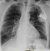
Case report: A 63-year-old white man with chronic obstructive pulmonary disease (COPD) was found to have a large pleural mass on chest radiography during a pre-operative assessment. The tumor was biopsied and findings were consistent with solitary fibrous tumor of the pleura.
Conclusions: SFTPs are generally considered benign tumors although there is a risk of malignant transformation and recurrence. Imaging studies play an important role in identifying the tumor and planes of resection, and histologic diagnosis is critical in differentiating SFTP from other type of pleural masses. Surgical resection is main therapy of choice.
Figures


References
-
- Chan JK. Solitary fibrous tumour: everywhere, and a diagnosis in vogue. Histopathology. 1997;31:568–76. - PubMed
-
- De Perrot M, Fischer S, Brundler MA, et al. Solitary fibrous tumors of the pleura. Ann Thorac Surg. 2002;74:285–93. - PubMed
-
- Sung SH, Chang JW, Kim J, et al. Solitary fibrous tumors of the pleura: surgical outcome and clinical course. Ann Thorac Surg. 2005;79:303–7. - PubMed
-
- Zellos LS, Sugarbaker DJ. Multimodality treatment of diffuse malignant pleural mesothelioma. Semin Oncol. 2002;29:41–50. - PubMed
-
- Dalton WT, Zolliker AS, McCaughey WT, et al. Localized primary tumors of the pleura: an analysis of 40 cases. Cancer. 1979;44:1465–75. - PubMed
Publication types
MeSH terms
LinkOut - more resources
Full Text Sources

