

Roentgenographic Evaluation of the Spine in Patients With Osteogenesis Imperfecta
- PMID: 26632680
- PMCID: PMC5058949
- DOI: 10.1097/MD.0000000000001841
Roentgenographic Evaluation of the Spine in Patients With Osteogenesis Imperfecta
Abstract
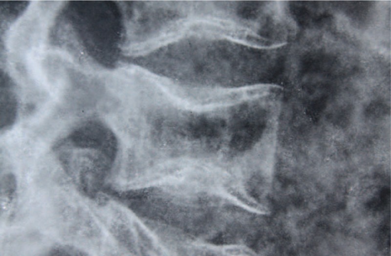
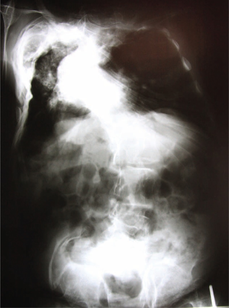
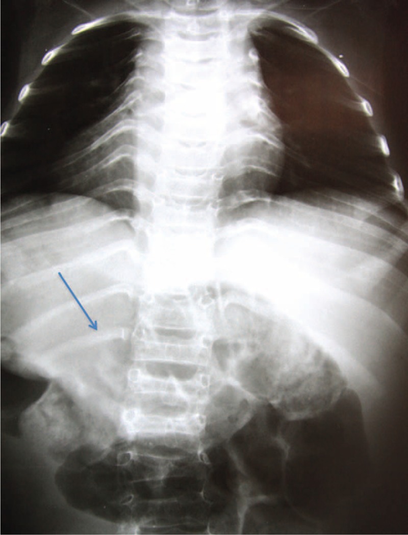
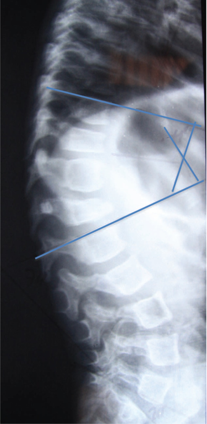
Osteogenesis imperfecta (OI) is a hereditary connective tissue disorder that leads to bone weakness and deformities, especially in the spine, which can lead to poor outcomes.The aim of this study was to find patterns and risk factors in spinal deformities in patients with OI.In a retrospective study, 70 patients with OI were selected. Radiographs of the spine were evaluated. We observed the presence or absence of the following changes: biconcave vertebrae, chest and vertebral deformities, unilateral rib, and thoracolumbar kyphosis. The greater curve was considered the primary one, and the secondary curve considered compensatory.In the study sample, we observed that the patients' ages ranged between 7 and 50 years, with a mean equal to 13 years, and 76% had scoliosis. In 68% of cases the main curve in the thoracic region was observed with the convexity to the right.The following was found in patients with OI: scoliosis, biconcave vertebrae, vertebral and chest deformity, unilateral rib, and thoracolumbar kyphosis. The thoracolumbar kyphosis is highly associated with thoracic hypokyphosis in patients with OI.
Conflict of interest statement
The authors have no funding and conflicts of interest to disclose.
Figures
References
-
- Byers PH, Steiner RD. Osteogenesis imperfect. Annu Rev Med 1992; 43:269–282. - PubMed
-
- Vetter U, Pontz B, Zauner E, et al. Osteogenesis imperfecta: a clinical study of the first ten years of life. Calcif Tissue Int 1992; 50:36–41. - PubMed
-
- Ishikawa S, Kumar SJ, Takahashi HE, et al. Vertebral body shape as a predictor of spinal deformity in osteogenesis imperfect. J Bone Joint Surg Am 1996; 78:212–219. - PubMed
-
- Shands A, Eisberg H. The incidence of scoliosis in the state of Delaware; a study of 50,000 minifilms of the chest made during a survey for tuberculosis. J Bone Joint Surg Am 1995; 37:1243–1249. - PubMed
MeSH terms
LinkOut - more resources
Full Text Sources
Medical
