

STROBE--Radiation Ulcer: An Overlooked Complication of Fluoroscopic Intervention: A Cross-Sectional Study
- PMID: 26632903
- PMCID: PMC4674206
- DOI: 10.1097/MD.0000000000002178
STROBE--Radiation Ulcer: An Overlooked Complication of Fluoroscopic Intervention: A Cross-Sectional Study
Abstract
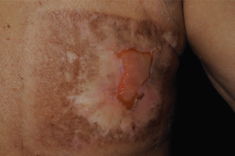
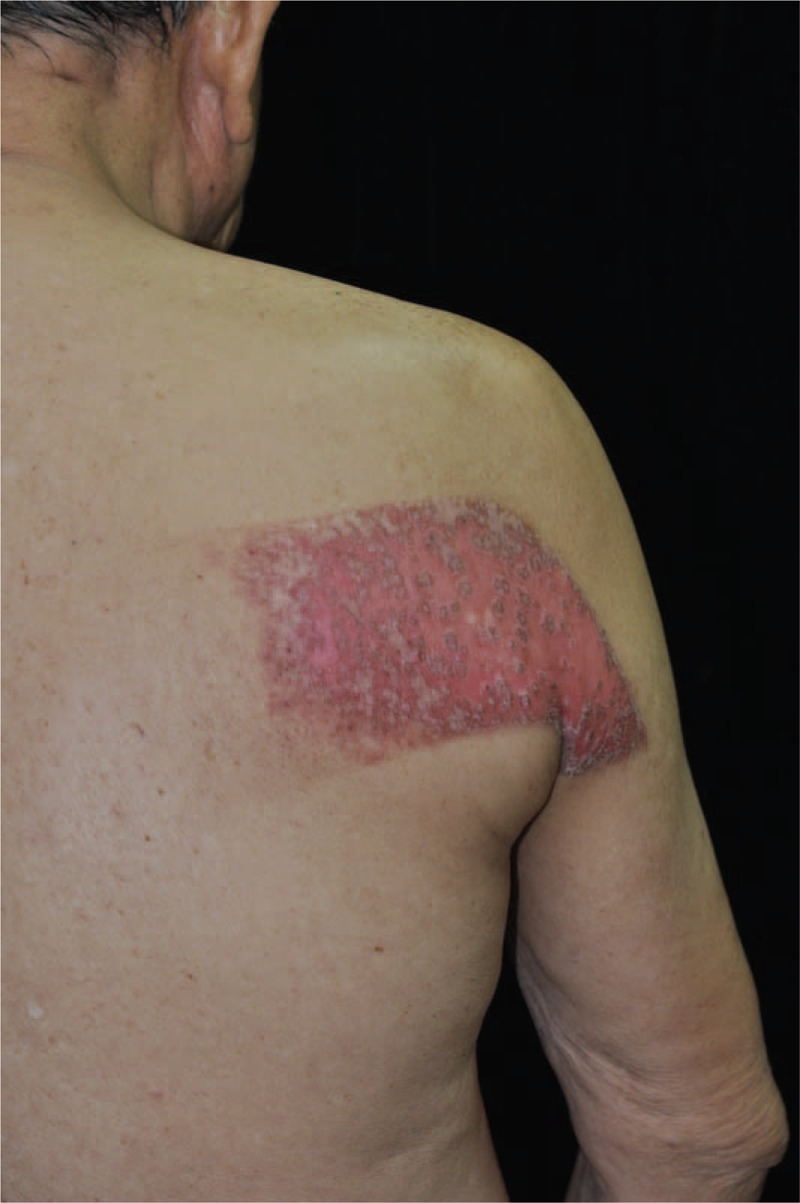
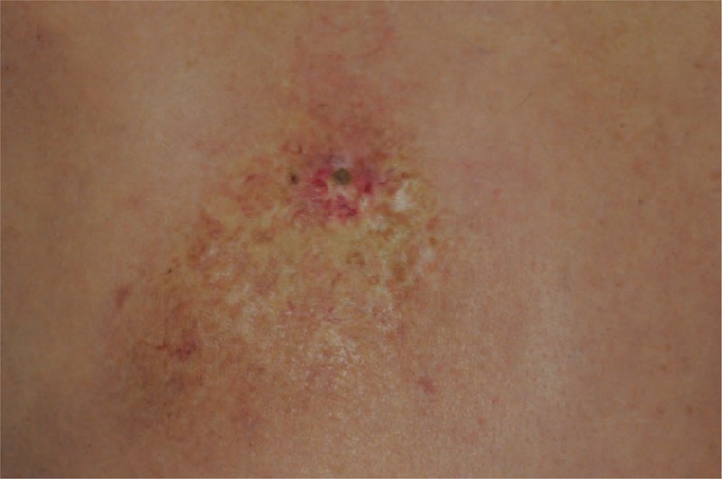
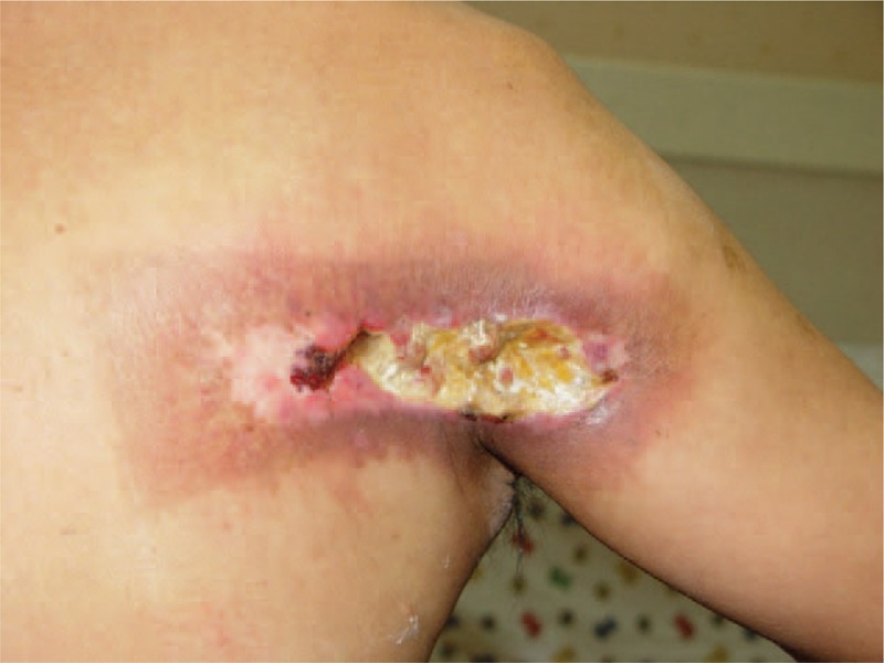
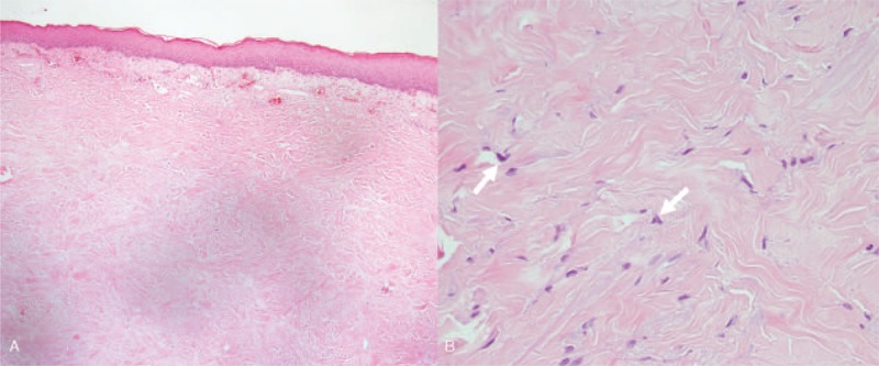
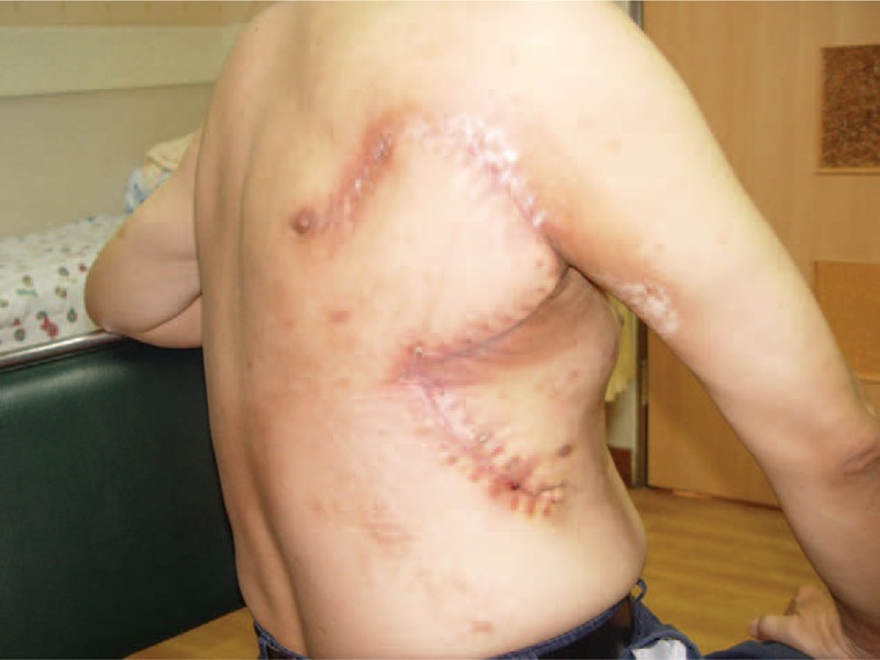
With increasing numbers of percutaneous coronary intervention (PCI) and complex cardiac procedures, higher accumulated radiation dose in patient has been observed. We speculate cardiac catheter intervention induced radiation skin damage is no longer rare.To study the incidence of cardiac fluoroscopic intervention induced radiation ulcer. We retrospectively reviewed medical records of those who received cardiac fluoroscopic intervention in our hospital during 2012 to 2013 for any events of radiation ulcer. Only patients, whose clinical photos were available for reviewing, would be included for further evaluation. The diagnosis of radiation ulcers were made when there is a history of PCI with pictures proven skin ulcers, which presented typical characteristics of radiation injury. Nine patients with radiation ulcer were identified and the incidence was 0.34% (9/2570) per practice and 0.42% (9/2124) per patient. Prolonged procedure time, cumulative multiple procedures, right coronary artery occlusion with chronic total occlusion, obesity, and diabetes are frequent characteristics. The onset interval between the first skin manifestation and the latest radiation exposure varied from 3 weeks to 3 months. The histopathology studies failed to make diagnosis correctly in 5 out of 6 patients. To make thing worse, skin biopsy exacerbated the preexisting radiation dermatitis. Notably, all radiation ulcers were refractory to conventional wound care. Surgical intervention was necessary to heal the wound. Diagnosis of cardiac fluoroscopy intervention induced radiation skin damage is challenging and needs high index of clinical suspicion. Minimizing the radiation exposure by using new approaches is the most important way to prevent this complication. Patient education and a routine postprocedure dermatology follow up are mandatory in high-risk groups for both radiation skin damage and malignancies. This is a retrospective study, thus the true incidence of radiation ulcer caused by cardiac fluoroscopic intervention could be higher.
Conflict of interest statement
The authors have no funding and conflicts of interest to disclose.
Figures
References
-
- Aerts A, Decraene T, van den Oord JJ, et al. Chronic radiodermatitis following percutaneous coronary interventions: a report of two cases. J Eur Acad Dermatol Venereol 2003; 17:340–343. - PubMed
-
- Khouzam RN, Soufi MK, Nakhla R, et al. Outpatient percutaneous coronary intervention: has its time come? J Invasive Cardiol 2014; 26:E167–E169. - PubMed
-
- Mozaffarian D, Benjamin EJ, Go AS, et al. Heart disease and stroke statistics—2015 update: a report from the American Heart Association. Circulation 2015; 131:e29–e322. - PubMed
-
- Brilakis E. Manual of Coronary Chronic Total Occlusion Interventions: A Step-by-Step Approach. Waltham, MA: Elsevier; 2013.
MeSH terms
LinkOut - more resources
Full Text Sources
Miscellaneous
