. 2015 Dec 1;7(12):6233-40.
doi: 10.3390/v7122934.

Identification of a New Ribonucleoside Inhibitor of Ebola Virus Replication

Affiliations

Identification of a New Ribonucleoside Inhibitor of Ebola Virus Replication

Olivier Reynard et al. Viruses. .

Erratum in

Abstract

The current outbreak of Ebola virus (EBOV) in West Africa has claimed the lives of more than 15,000 people and highlights an urgent need for therapeutics capable of preventing virus replication. In this study we screened known nucleoside analogues for their ability to interfere with EBOV replication. Among them, the cytidine analogue β-d-N4-hydroxycytidine (NHC) demonstrated potent inhibitory activities against EBOV replication and spread at non-cytotoxic concentrations. Thus, NHC constitutes an interesting candidate for the development of a suitable drug treatment against EBOV.

Keywords: Ebola; Filovirus; Marburg; antiviral; inhibitors; polymerase.


Figures

Figure 1
Figure 1
Screening of antiviral compounds against Ebola virus (EBOV). Vero E6 cells were infected with EBOV expressing GFP (EBOV/GFP) at an multiplicity of infection (MOI) of 0.1 in the presence of different chemical compounds (see Table 1) at the indicated concentration. Forty-eight hours afterwards, cells were detached from the plate with trypsin and fixed for 20 min with 3% PBS buffered paraformaldehyde prior to flow cytometry analysis on a Beckman Coulter Gallios apparatus (Beckman, Brea, CA, USA). Neg value indicate the background level in the non infected condition. The graph presents both the % of GFP-positive cells, reflecting the extent of viral spread and the median fluorescence intensity (MFI), simulating the level of viral transcription. All data represents averages and SEM of three independent experiments. (Please replace by: All data represents averages and SD of three independent experiments. * A student test was performed between value of compound 9 and the Mock treated condition, p-value below 0.05. The data are representative of the values of two different experiments performed in triplicate).
Figure 2
Figure 2
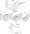
Dose-response assessment and β-D-N4-hydroxycytidine (NHC) cytotoxicity Vero E6 cells (A) and monocyte-derived macrophages (B) were infected with EBOV/GFP at an MOI of 0.1, and maintained in the presence of NHC at the indicated concentrations. The cells were analyzed forty-eight hours post infection as described in Figure 1. Macrophages were differentiated upon incubation of monocytes (three different donors) with 100 ng/mL of M-CSF (EuroBio, Courtaboeuf, France). The graph displays both the percentage of infected cells and the GFP MFI as measured by flow cytometry (Beckman Coulter, Gallios, Brea, CA, USA); (C) MTT (3-(4,5-dimethylthiazol-2-yl)-2,5-diphenyltetrazolium bromide) cytotoxicity assay. Vero E6 cells and monocyte-derived macrophages were maintained in the NHC presence for 48 h prior to addition of MTT (5 µg/mL, Sigma Aldrich, Saint-Quentin Fallavier, France) followed by 3 h incubation and optical density analysis The graph displays the OD value normalized to the 100% cytotoxicity control.
Figure 3
Figure 3
Mode of NHC antiviral activity: (A) NHC effect on expression of GFP. Vero E6 cells were transfected with 3 µg of plasmid DNA expressing GFP (peGFP N1, Clontech) using FugeneHD (Promega), GFP MFI was measured on a Becton Dickinson, LSRII. The graph presents averages and SEM of three independent experiments; (B) qRT-PCR analysis of genomic and anti-genomic RNA. Vero E6 cells were infected with EBOV/GFP at an MOI of 0.1 and analyzed 24 h post infection by qRT-PCR. The reverse transcription Iscript select cDNA synthesis (Biorad) was followed by qPCR (Faststart universal Sybr green and LC96 apparatus, both Roche diagnostics) using forward and reverse primer in the non-transcribed trailer region (TR-F: 5’ATTACTGCCGCAATGAATTTAACGC (18429), TR-R: 5’AACAATATGAGCCCAGACCTTTCG (18517)). To distinguish between genomic and antigenomic RNAs, the reverse transcription step was carried out using either a forward or a reverse primer, respectively. The data are representative of the values of two separate experiments performed in duplicate. Data were normalized by using GAPDH Fw and Rev primer (5′CACCCACTCCTCCACGTTTGAC and 5′GTCCACCACCCTGTTGCTGTAG respectively); (C) Inhibition of EBOV replication and spread by NHC. Vero E6 cells were infected with EBOV/GFP at an MOI of 0.01 and were analyzed daily by flow cytometry. Left panel: present of infected cells; right panel: relative GFP fluorescent intensity. The data are representative of the values of two different experiments performed in duplicate. * A student test was performed between value of treated and the Mock treated condition at day 4, p-value below 0.05 for both 1.9 and 7.2 µM; (D) Effect of NHC on Marburg virus replication. Vero E6 cells were infected with Marburg virus (Musoke isolate) at MOI 0.1 and treated with NHC (7.2 µM) for a period of four days prior to WB analysis. NI, non-Infected cells; I, infected cells; I-NHC, infected cells treated with NHC at day 0; I-NHC2, infected cells treated with NHC twice. Second treatment was performed at day 2 after cell washing and media replenishment. Western blot analysis of cells and culture supernatants was made using a mouse anti-Marburg GP1 antibody (clone 6589). The data are representative of the values of two different experiments.

References

    1. Incident Management System Ebola Epidemiology Team, CDC. Guinea Interministerial Committee for Response Against the Ebola Virus. World Health Organization. CDC Guinea Response Team. Liberia Ministry of Health and Social Wealfare. CDC Liberia Response Team. Sierra Leone Ministry of Health and Sanitation. CDC Sierria Leone Response Team. Viral Special Pathogens Branch, National Center for Emerging and Zoonotic Infectious Diseases, CDC. Centers for Disease Control and Prevention (CDC) Update: Ebola virus disease epidemic—West Africa, February 2015. MMWR Morb. Mortal. Wkly. Rep. 2015;64:186–187. - PMC - PubMed
    1. Kuhn J.H., Becker S., Ebihara H., Geisbert T.W., Johnson K.M., Kawaoka Y., Lipkin W.I., Negredo A.I., Netesov S.V., Nichol S.T., et al. Proposal for a revised taxonomy of the family filoviridae: Classification, names of taxa and viruses, and virus abbreviations. Arch. Virol. 2010;155:2083–2103. doi: 10.1007/s00705-010-0814-x. - DOI - PMC - PubMed
    1. Feldmann H., Klenk H.D., Sanchez A. Molecular biology and evolution of filoviruses. Arch. Virol. Suppl. 1993;7:81–100. - PubMed
    1. Volchkov V.E., Volchkova V.A., Slenczka W., Klenk H.D., Feldmann H. Release of viral glycoproteins during Ebola virus infection. Virology. 1998;245:110–119. doi: 10.1006/viro.1998.9143. - DOI - PubMed
    1. Volchkova V.A., Feldmann H., Klenk H.D., Volchkov V.E. The nonstructural small glycoprotein sGP of Ebola virus is secreted as an antiparallel-orientated homodimer. Virology. 1998;250:408–414. doi: 10.1006/viro.1998.9389. - DOI - PubMed

Publication types