. 2015 Dec 22;113(12):1720-9.
doi: 10.1038/bjc.2015.367. Epub 2015 Dec 3.

FAT4 functions as a tumour suppressor in gastric cancer by modulating Wnt/β-catenin signalling

Affiliations

FAT4 functions as a tumour suppressor in gastric cancer by modulating Wnt/β-catenin signalling

Jian Cai et al. Br J Cancer. .

Abstract

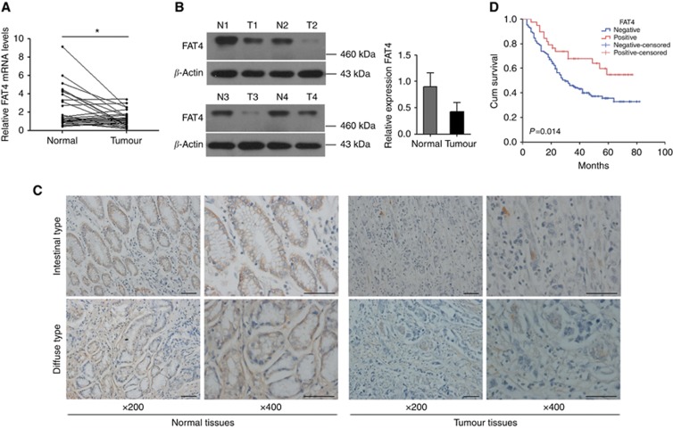
Background: FAT4, a cadherin-related protein, was shown to function as a tumour suppressor; however, its role in human gastric cancer remains largely unknown. Here, we investigated the role of FAT4 in gastric cancer and examined the underlying molecular mechanisms.

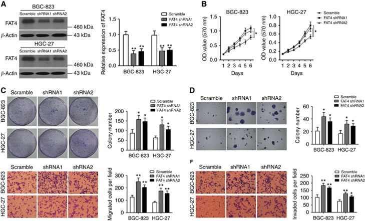
Methods: The expression of FAT4 was evaluated by immunohistochemistry, western blotting, and qRT-PCR in relation to the clinicopathological characteristics of gastric cancer patients. The effects of FAT4 silencing on cell proliferation, migration, and invasion were assessed by the MTT (3-(4,5-dimethylthiazol-2-yl)-2,5-diphenyltetrazolium) assay, and migration and invasion assays in gastric cancer cell lines in vitro and in a mouse xenograft model in vivo.

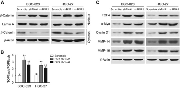
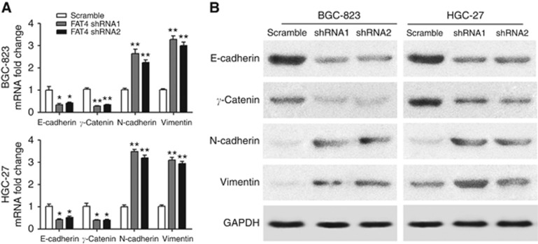
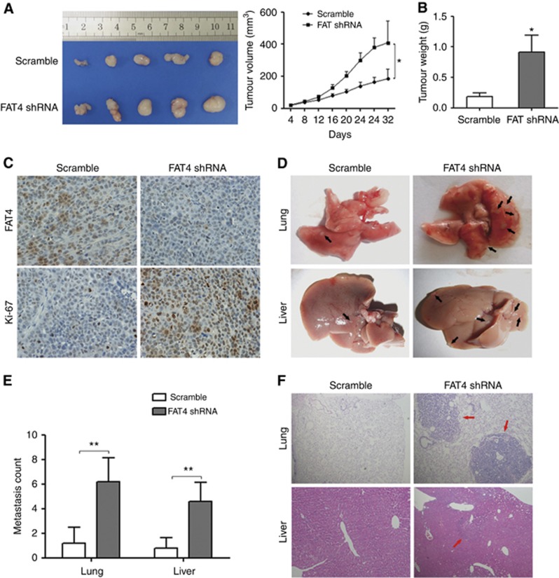
Results: Downregulation of FAT4 expression in gastric cancer tissues compared with adjacent normal tissues was correlated with lymph-node metastasis and poor survival. Knockdown of FAT4 promoted the growth and invasion of gastric cancer cells via the activation of Wnt/β-catenin signalling, and induced epithelial-to-mesenchymal transition (EMT) in gastric cancer cells, as demonstrated by the upregulation and downregulation of mesenchymal and epithelial markers. Silencing of FAT4 promoted tumour growth and metastasis in a gastric cancer xenograft model in vivo.

Conclusions: FAT4 has a tumour suppressor role mediated by the modulation of Wnt/β-catenin signalling, providing potential novel targets for the treatment of gastric cancer.


Figures

Figure 1
Figure 1
FAT4 expression in gastric cancer tissues. (A) FAT4 mRNA expression levels in 30 paired gastric cancer tissues and adjacent non-cancerous tissues expressed as relative expression normalised to the expression of β-actin and calculated by the 2−ΔΔct method. *P<0.05 by Mann–Whitney U-test. (B) FAT4 protein expression in representative samples of gastric cancer tissues (T) and paired adjacent non-cancerous tissues (N). *P<0.05 by Student's t-test. (C) Immunohistochemical staining of FAT4 in gastric cancer tissues. Original magnification, × 200, × 400; bar=100 μm. (D) Kaplan–Meier survival analysis according to FAT4 expression in gastric cancer patients (N=160). The P-values from log-rank tests comparing the two KM curves are shown.
Figure 2
Figure 2
FAT4 knockdown promotes the proliferation and invasion of BGC-823 and HGC-27 cells. BGC-823 and HGC-27 cells were infected with FAT4 shRNA or scramble control shRNA. FAT4 protein expression was analysed by western blotting and quantified by densitometric scanning (A). Cell proliferation was measured by MTT assay (B). Colony formation was analysed by colony-formation (C) and soft agar (D) assays. Migration (E) and invasion (F) were analysed in BGC-823 and HGC-27 cells infected as indicated. Representative images of colony formation, migrating, and invading cells in scramble and FAT4-silenced gastric cancer cells are shown. *P<0.05, **P<0.01 by Student's t-test.
Figure 3
Figure 3
Effect of FAT4 knockdown on Wnt/β-catenin signalling. (A) Altered nuclear translocation of β-catenin in response to FAT4 knockdown. Nuclear and cytosolic extracts of BGC-823 and HGC-27 cells were analysed by western blotting with Lamin A and β-actin as a loading control. (B) Cells were transfected with TOPflash or FOPflash and Renilla pRL-TK plasmids and subjected to dual-luciferase assays 48 h after transfection. Reporter activity was normalised to Renilla luciferase activity. (C) Western blot analysis of Wnt target gene expression in response to FAT4 knockdown in BGC-823 and HGC-27 cells. β-Actin was used as the loading control. **P<0.01 by Student's t-test.
Figure 4
Figure 4
FAT4 knockdown-induced gastric cancer cell growth, invasion, and migration are mediated by the Wnt/β-catenin signalling pathway. BGC-823 and HGC-27 cells were transfected with or without shRNA (shRNA1) against FAT4 and siRNA targeting β-catenin, and analysed by western blotting (A), MTT assay (B), colony-formation assay (C), and migration and invasion assays (D and E). *P<0.05, **P<0.01 by Student's t-test.
Figure 5
Figure 5
FAT4 modulates the expression of EMT markers. BGC-823 and HGC-27 cells were infected with scramble control or shRNAs against FAT4 and the mRNA (A) and protein (B) expression of EMT markers was determined by RT–PCR and western blotting, respectively. *P<0.05, **P<0.01 by Student's t-test.
Figure 6
Figure 6
Effect of FAT4 knockdown on tumorigenesis and metastasis in vivo. (A) Photographs of excised tumours and tumour growth curves in mice transplanted with BGC-823/Scramble or BGC-823/FAT4 shRNA cells (FAT4 shRNA1) (N=5 per group). (B) After 32 days, the mice were killed and tumours were weighed. (C) IHC staining for FAT4 and Ki-67 in sections of transplanted tumours. (D) BGC-823/Scramble and BGC-823/FAT4 shRNA cells (6 × 10⁶ cells per mouse) were injected into the tail vein of two groups of nude mice. Four weeks post injection, the mice were killed and the lungs and livers were removed and paraffin embedded. Arrows indicate surface metastatic nodules. (E) Number of visible surface metastatic lesions on the lungs and livers of individual mice (N=5 per group) receiving an orthotopic injection of the indicated cells. (F) Representative images of haematoxylin and eosin-stained lung and liver sections of mice injected with the indicated cells. *P<0.05, **P<0.01 by Student's t-test.

References

    1. Berx G, van Roy F (2009) Involvement of members of the cadherin superfamily in cancer. Cold Spring Harb Perspect Biol 1: a003129. - PMC - PubMed
    1. Bossuyt W, Chen CL, Chen Q, Sudol M, McNeill H, Pan D, Kopp A, Halder G (2014) An evolutionary shift in the regulation of the Hippo pathway between mice and flies. Oncogene 33: 1218–1228. - PMC - PubMed
    1. Braun GS, Kretzler M, Heider T, Floege J, Holzman LB, Kriz W, Moeller MJ (2007) Differentially spliced isoforms of FAT1 are asymmetrically distributed within migrating cells. J Biol Chem 282: 22823–22833. - PubMed
    1. Catalano V, Labianca R, Beretta GD, Gatta G, de Braud F, Van Cutsem E (2009) Gastric cancer. Crit Rev Oncol Hematol 71: 127–164. - PubMed
    1. Clements WM, Wang J, Sarniak A, Kim OJ, MacDonald J, Fenoglio-Preiser C, Groden J, Lowy AM (2002) β-Catenin mutation is a frequent cause of Wnt pathway activation in gastric cancer. Cancer Res 62: 3503–3506. - PubMed

Publication types

MeSH terms