. 2015 Dec 1:5:44.
doi: 10.1186/s13395-015-0070-6. eCollection 2015.

Elusive sources of variability of dystrophin rescue by exon skipping

Affiliations

Elusive sources of variability of dystrophin rescue by exon skipping

Maria Candida Vila et al. Skelet Muscle. .

Abstract

Background: Systemic delivery of anti-sense oligonucleotides to Duchenne muscular dystrophy (DMD) patients to induce de novo dystrophin protein expression in muscle (exon skipping) is a promising therapy. Treatment with Phosphorodiamidate morpholino oligomers (PMO) lead to shorter de novo dystrophin protein in both animal models and DMD boys who otherwise lack dystrophin; however, restoration of dystrophin has been observed to be highly variable. Understanding the factors causing highly variable induction of dystrophin expression in pre-clinical models would likely lead to more effective means of exon skipping in both pre-clinical studies and human clinical trials.

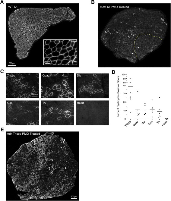
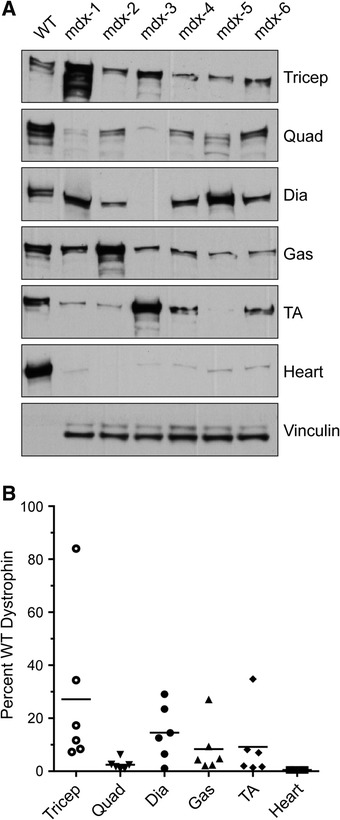
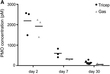
Methods: In the present study, we investigated possible factors that might lead to the variable success of exon skipping using morpholino drugs in the mdx mouse model. We tested whether specific muscle groups or fiber types showed better success than others and also correlated residual PMO concentration in muscle with the amount of de novo dystrophin protein 1 month after a single high-dose morpholino injection (800 mg/kg). We compared the results from six muscle groups using three different methods of dystrophin quantification: immunostaining, immunoblotting, and mass spectrometry assays.

Results: The triceps muscle showed the greatest degree of rescue (average 38±28 % by immunostaining). All three dystrophin detection methods were generally concordant for all muscles. We show that dystrophin rescue occurs in a sporadic patchy pattern with high geographic variability across muscle sections. We did not find a correlation between residual morpholino drug in muscle tissue and the degree of dystrophin expression.

Conclusions: While we found some evidence of muscle group enhancement and successful rescue, our data also suggest that other yet-undefined factors may underlie the observed variability in the success of exon skipping. Our study highlights the challenges associated with quantifying dystrophin in clinical trials where a single small muscle biopsy is taken from a DMD patient.

Keywords: Duchenne muscular dystrophy; Dystrophin; Exon skipping; Variability; mdx-23.


Figures

Fig. 1
Fig. 1
Variability of dystrophin protein expression, as shown by IF after PMO injection. a Representative images of C57BL/10 (WT) and b PMO-treated mdx tibialis anterior sections stained for dystrophin. The WT control shows uniform IF staining for dystrophin. Insert at ×40 shows expected staining pattern for dystrophin-positive fibers. b PMO-treated mdx tibialis anterior shows a mosaic staining pattern and clustering of positive fibers. The yellow line represents the border between the tibialis anterior and EDL. Quantification was performed on the entire area of the muscle section. c Representative images of mouse mdx-6 showing variability between the muscles of the same animal. Images were selected to show positive fiber clustering and do not represent total area quantification. d IF quantification of diaphragm, gastrocnemius, heart, quadriceps, tibialis anterior, and triceps for all mice (n = 6). e Geographic variability observed within the highly rescued triceps from mouse mdx-1. All tissues were sectioned (10-μm thick), stained, and probed with goat anti-rabbit IgG Alexa 594 antibody. Dystrophin-positive fibers were normalized to the area of the muscle section and the WT percentage of positive fibers. Original magnification for a, b, e = ×20; scale bar, 500 μm; for c = ×40; scale bar, 100 μm
Fig. 2
Fig. 2
Dystrophin protein expression as detected by IB 1 month after PMO injection. a IB of protein lysates from diaphragm, heart, gastrocnemius, quadriceps, tibialis anterior, and triceps in PMO-treated mdx mice (n = 6) vs. WT control shows variability of dystrophin expression between muscles in PMO-injected mdx mice. There is variation in the same muscles between different mice (across) and different muscles in the same individual mouse (down). WT samples were serially diluted to 3.125 μg for protein loading. Vinculin (117 kDa) was used as a loading control. Densitometric analysis was performed using Quantity One software. b Dystrophin quantification by IB, demonstrating the percentage dystrophin expression in PMO-injected mice vs. WT (set to 100 %). Plots show high variability between mice within a muscle group and between muscles. All the data are presented as mean percentages
Fig. 3
Fig. 3
Dystrophin protein expression as detected by MS. a Triceps, tibialis anterior, and gastrocnemius RIPA buffer extracts were analyzed by MS. The percentage of dystrophin protein expression in PMO-injected mdx mice was compared to the dystrophin percentage in WT. All data are presented as mean percentages
Fig. 4
Fig. 4
De novo dystrophin restoration varies independently of muscle/fiber type. a IB of the predominately slow-twitch muscle soleus and b fast-twitch EDL muscle shows no preference for dystrophin rescue by muscle fiber type. WT samples were loaded at 25 μg, and vinculin was used as a loading control. For mdx mice, 75 μg of total protein was loaded. c IF in serial sections from the triceps muscle that showed the highest dystrophin rescue for fiber-type identification of type 1, type 2a, type 2b, and embryonic myosin heavy chain (eMHC) isoforms (green) and dystrophin (red). Asterisks and pound signs indicate the same muscle fiber in different images. Original magnification for a = ×40; scale bar, 100 μm
Fig. 5
Fig. 5
Correlation between residual PMO in muscle and de novo dystrophin. a–c Percentage of WT dystrophin as measured by a IF, and PMO levels measured by hybridization ELISA were plotted with a regression line. The same analysis was performed for dystrophin quantified by b IB and c MS. There was no significant correlation, as illustrated by the regression lines. Note that for MS we have fewer data points because we measured only three muscle groups by this method (N = 18)
Fig. 6
Fig. 6
Time course of residual PMO concentration. a Residual morpholino concentration was measured in the triceps and gastrocnemius muscle extracts at 2 (n = 3), 7 (n = 3), and 30 (n = 6) days after PMO administration. No statistically significant differences were found between the concentrations of PMO in the two muscle groups for a given time point. We observed a decline in the PMO concentration in muscle over time

References

    1. Hoffman EP. Genotype/phenotype correlations in Duchenne/Becker dystrophy. Mol Cell Biol Hum Dis Serv. 1993;3:12–36. - PubMed
    1. Hoffman EP, Monaco AP, Feener CC, Kunkel LM. Conservation of the Duchenne muscular dystrophy gene in mice and humans. Science. 1987;238(4825):347–50. doi: 10.1126/science.3659917. - DOI - PubMed
    1. Hoffman EP, Schwartz L. Dystrophin and disease. Mol Aspects Med. 1991;12(3):175–94. doi: 10.1016/0098-2997(91)90001-3. - DOI - PubMed
    1. Roses AD, Herbstreith MH, Appel SH. Membrane protein kinase alteration in Duchenne muscular dystrophy. Nature. 1975;254(5498):350–1. doi: 10.1038/254350a0. - DOI - PubMed
    1. Hutter OF, Burton FL, Bovell DL. Mechanical properties of normal and mdx mouse sarcolemma: bearing on function of dystrophin. J Muscle Res Cell Motil. 1991;12(6):585–9. doi: 10.1007/BF01738447. - DOI - PubMed