. 2015 Dec 4:6:10038.
doi: 10.1038/ncomms10038.

Vinculin controls talin engagement with the actomyosin machinery

Affiliations

Vinculin controls talin engagement with the actomyosin machinery

Paul Atherton et al. Nat Commun. .

Abstract

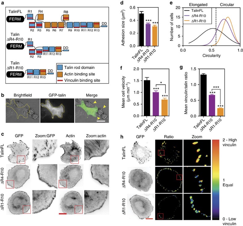
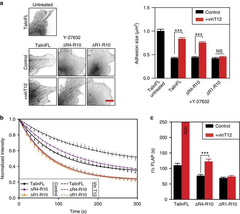
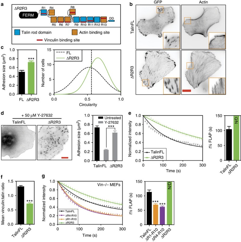
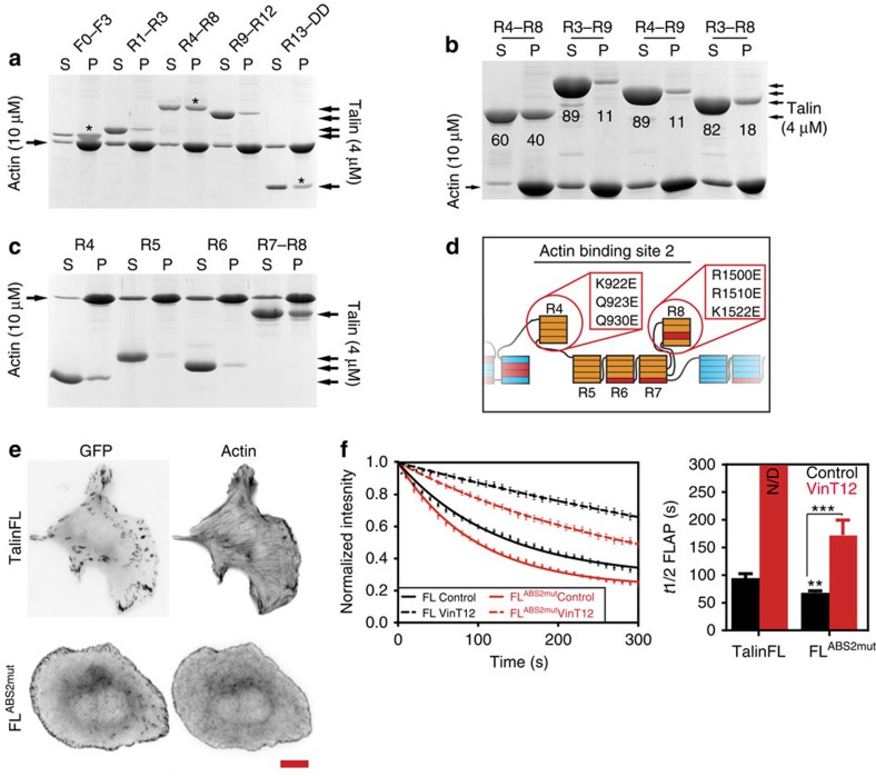
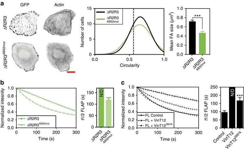
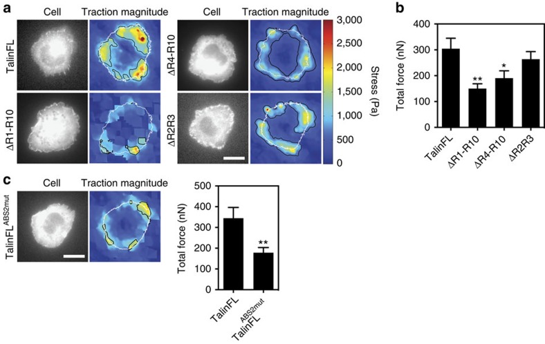
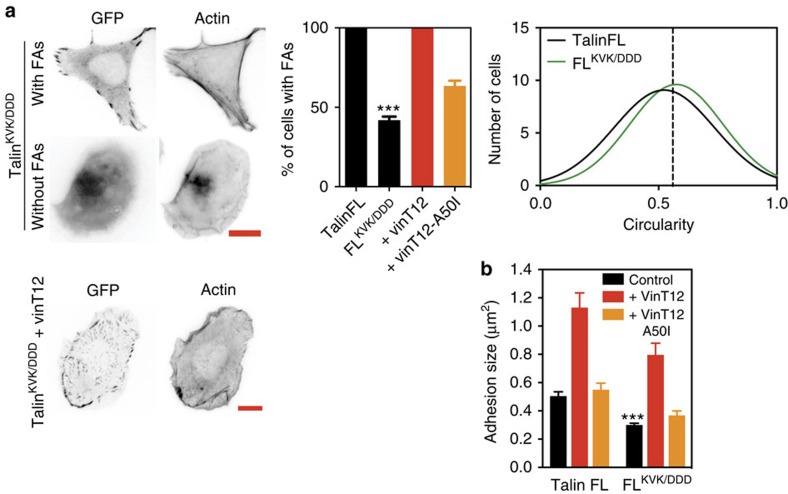
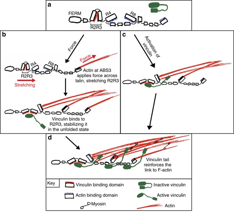
The link between extracellular-matrix-bound integrins and intracellular F-actin is essential for cell spreading and migration. Here, we demonstrate how the actin-binding proteins talin and vinculin cooperate to provide this link. By expressing structure-based talin mutants in talin null cells, we show that while the C-terminal actin-binding site (ABS3) in talin is required for adhesion complex assembly, the central ABS2 is essential for focal adhesion (FA) maturation. Thus, although ABS2 mutants support cell spreading, the cells lack FAs, fail to polarize and exert reduced force on the surrounding matrix. ABS2 is inhibited by the preceding mechanosensitive vinculin-binding R3 domain, and deletion of R2R3 or expression of constitutively active vinculin generates stable force-independent FAs, although cell polarity is compromised. Our data suggest a model whereby force acting on integrin-talin complexes via ABS3 promotes R3 unfolding and vinculin binding, activating ABS2 and locking talin into an actin-binding configuration that stabilizes FAs.


Figures

Figure 1
Figure 1. Talin rod domains regulate cell polarity and migration, FA morphology and FA composition.
(a) Cartoon of talin constructs expressed as N-terminally tagged GFP-fusion proteins. The talin FERM domain is linked to the flexible talin rod which consists of 13 helical bundles (R1-R13) terminating in a dimerization helix (DD). Constructs in which the R4-R10 (talΔR4-R10) and R1-R10 (talΔR1-R10) domains have been deleted are shown. Colours indicate binding sites for actin (orange) and vinculin (red). (b) Talin1 and talin2 knockout (TKO) cells cells transfected with talinFL, 24 h after plating on fibronectin; arrows indicate non-transfected cells, which do not spread. (c) TKO cells expressing indicated GFP-talin fusion constructs were plated on fibronectin and stained for F-actin. Magnified regions are from the area framed in red. Note the colocalisation of talΔR1-R10 or talΔR4-R10 with F-actin at the cell edge. Scale bars, 10 μm. (d) Quantification of FA size in TKO cells expressing indicated constructs. Note that talΔR1-R10 or talΔR4-R10 have smaller FAs compared with cells expressing talinFL (n>70 cells, from three independent experiments, error bars are ±s.e.m., ***=P<0.001 (ANOVA)). (e) Quantification of cell circularity of cells expressing GFP-talin constructs. Note that cells expressing talΔR1-R10 or talΔR4-R10 are more circular than cells expressing talinFL (n=90 cells, from three independent experiments). (f) TKO cells expressing the three GFP-talin constructs were tracked over 24 h. Both talΔR1-R10 and talΔR4-R10 cells have significantly reduced velocity compared with talinFL cells; talΔR1-R10 cells are slower than talΔR4-R10 cells (n=20 cells, from two independent experiments, error bars are±s.e.m., *=P<0.05; ***=P<0.001 (ANOVA). (g,h) Ratiometric imaging was used to determine the proportion of vinculin present at adhesions in TKO cells expressing indicated constructs. Quantitative analysis (in g) shows that vinculin levels are reduced in adhesions in talin rod deletion mutants. Cells expressing the talΔR1-R10 construct (the largest deletion) had the lowest levels of vinculin in adhesion complexes. Scale bar in h, 10 μm. (n>65 cells, from three independent experiments, error bars are±s.e.m., ***=P<0.001 (ANOVA) compared with talinFL unless where indicated).
Figure 2
Figure 2. Vinculin regulates talin by binding to R1-R3 domains.
(a) NIH3T3 cells expressing talinFL, talΔR1-R10 or talΔR4-R10 with vinFL (control) or vinT12 were treated with DMEM containing 50 μM Y-27632 or an equivalent volume of water for 30 min before fixation. The effect on adhesions was analysed assessing the signal from the expressed GFP-talin fusion constructs. FAs in control cells expressing talinFL are dramatically reduced in size following treatment with Y-27632. Note that talinFL and talΔR4-R10, but not talΔR1-R10, in FAs are stabilised by co-expression of vinT12 (n=15 cells, representative of three independent experiments, error bars are±s.e.m., ***=P<0.001 (ANOVA)); scale bar, 5 μm. (b) Curves from fluorescence loss after photoactivation (FLAP) experiments in NIH3T3 cells expressing either talinFL, talΔR4-R10 or talΔR1-R10 with and without vinT12. (c) Quantification of half-time of FLAP (t½ FLAP) to assess the mobility of talin constructs in FAs. Note that vinT12 reduces the mobility (increase of t½) of talinFL and talΔR4-R10 but not talΔR1-R10. The almost linear decay of talinFL in the presence of vinT12 prevented appropriate fitting, hence accurate t½ FLAP could not be determined; N/D, not determined (n=25–66 FAs, from three independent experiments, error bars are±s.e.m., ***=P<0.001 (ANOVA)).
Figure 3
Figure 3. Deletion of Talin R2R3 stabilises FAs.
(a) Cartoon of the talΔR2R3 construct. (b,c) TKO cells expressing talΔR2R3 have more prominent actin stress fibres associated with larger FAs than those in cells expressing talinFL; cells expressing talΔR2R3 are more circular than talinFL cells (n=70–90 cells, from three independent experiments, error bars are±s.e.m., ***=P<0.001 (ANOVA)), scale bar in b, 10 μm. (d) Left panel, representative images of TKO cells expressing indicated constructs treated with DMEM containing 50 μM Y-27632 or an equivalent volume of water for 30 min. Note that Y-27632 resulted in a reduction of adhesion size of cells expressing talinFL compared with either untreated control cells or cells expressing talΔR2R3. (e) FLAP measurement curves of talΔR2R3 and talinFL in TKO cells. Curves (left panel) and quantification of t½ (right panel) show the reduced mobility of talΔR2R3 in comparison to talinFL (n=49–51 FAs, from three independent experiments). The almost linear decay of talΔR2R3 prevented appropriate fitting, hence accurate t½ FLAP could not be determined; N/D, not determined. (f) Quantification of vinculin recruitment in TKO cells expressing talΔR2R3. Note the reduced levels of vinculin in FAs of talΔR2R3 cells compared with levels in FAs of control cells expressing talinFL (n>70, from three independent experiments, error bars are±s.e.m., ***=P<0.001 (ANOVA)). (g) FLAP curves of experiments in MEFvin−/− cells (left panel) transfected with talinFL or talΔR2R3. Quantification of FLAP (right panel) showing t½ times for each construct. Note that even in absence of vinculin, talΔR2R3 has a slower turnover than talinFL. The almost linear decay of talΔR2R3 prevented appropriate fitting, hence accurate t½ FLAP could not be determined; N/D=not determined. (n=35–60 FAs, from three independent experiments, error bars are±s.e.m.).
Figure 4
Figure 4. ABS2 is a key regulator of talin.
(a,b) Recombinant talin polypeptides were incubated with F-actin, the actin pelleted, and supernatants (S) and pellets (P) analysed by SDS–PAGE. (a) Talin FERM domain F0-F3 (ABS1) followed by talin rod domains R1-R3, R4-R8 (ABS2), R9-R12 and R13-DD (ABS3). Asterisks show the domains containing the known ABSs. (b) Actin co-sedimentation assays show that actin binding to ABS2 is reduced when either domain R3 or R9 is present; numbers show percent band density (c) Actin co-sedimentation studies using the 4 sub-domains that make up ABS2; Note that R4 and R7-R8 bind actin. (d) Scheme outlining point mutations in the talin rod that reduce actin binding to ABS2. (e) Expression of talin ABS2 mutant (FLABS2mut) in TKO cells rescued cell spreading, but resulted in rounder cells with smaller FAs compared with talinFL cells (see also Supplementary Fig. 6a,b; scale bar, 10 μm). (f) FLAP experiments in TKO cells show that the turnover of talinFLABS2mut is increased compared with talinFL (FL control). Note that co-expression with vinT12 reduced the turnover of talinFLABS2mut to a lesser extent than talinFL. The almost linear decay of the talinFL in presence of vinT12 (FL VinT12) prevented appropriate fitting, hence accurate t½ FLAP could not be determined; N/D=not determined (n=28–56 FAs, from three independent experiments, error bars are±s.e.m., ***=P<0.001 (ANOVA)).
Figure 5
Figure 5. Talin R2R3 regulate the availability of ABS2 for actin binding.
(a) The talin ABS2 point mutations were introduced into talΔR2R3 and this construct (talΔR2R3ABS2mut) was expressed in TKO cells. Scale bar, 10 μm. TalΔR2R3ABS2mut cells had a similar circularity to talΔR2R3 cells, but lacked prominent actin stress fibres and had smaller FAs than talΔR2R3 cells (n>70 cells, from three independent experiments, error bars are±s.e.m., ***=P<0.001 (t-test)). (b) FLAP experiments show that talΔR2R3ABS2mut had a faster turnover than talΔR2R3 (n=48–51 FAs, from three independent experiments, error bars are±s.e.m.). (c) FLAP experiments of talinFL co-expressed with either vinT12 or a mutant form of vinT12 with reduced actin binding (vinT12I997A). Note that mutating the actin-binding site within the vinculin tail reduces the stabilizing effect of vinT12 (n=36–47 FAs, from three independent experiments, error bars are±s.e.m.).
Figure 6
Figure 6. The link between talin ABS2 and F-actin is critical for force exertion.
(a) Representative images from traction force microscopy (TFM) experiments. Colour spectrum indicates stress magnitude (Pa), with areas of low traction in blue and high traction in red. (b) Quantification of total force (nN) from TFM experiments shows that TKO cells expressing constructs lacking ABS2 (talΔR1-R10 and talΔR4-R10) exert less force than cells expressing talinFL (n=26–46 cells, error bars are±s.e.m., *=P<0.05, **=P<0.01 compared with talinFL (ANOVA)). (c) TFM experiments were repeated with the talinFLABS2mut construct. Cells expressing talinFLABS2mut had reduced force exertion compared with cells expressing talinFL (n=21–30 cells, error bars are±s.e.m., **=P<0.01 (t-test)). Scale bars in a and c, 10 μm.
Figure 7
Figure 7. The role of talin ABS3 in FA assembly can be bypassed by active vinculin.
(a) While expression of talinFLKVK/DDD in TKO cells rescues cell spreading,<50% of the cells display FAs connected with F-actin. FA formation in all cells was rescued by co-expression with vinT12 but not by a vinT12A50I mutant with reduced talin binding. Scale bars, 10 μm. (n>70 cells, ***=P<0.001 (t-test), error bars are±s.e.m.). (b) In those talinFLKVK/DDD cells that do have FAs, the FAs are smaller compared with those in talinFL cells. Co-expression with vinT12 (but not vinT12A50I) rescued FA size in cells expressing talinFLKVK/DDD (n>70 cells, ***=P<0.001 (ANOVA), error bars are±s.e.m.).
Figure 8
Figure 8. Model for vinculin supported talin engagement with the actomyosin machinery.
(a) Talin in which the vinculin and actin binding sites in R2R3 and R4-R8 (ABS2), respectively are cryptic. Inactive vinculin in green. (b,c) Two possible mechanisms for linking talin with the actomyosin machinery; (b) force-dependent pathway: actin binding to ABS3 leads to force exertion across talin, stretching R2R3 and unmasking its previously cryptic vinculin binding sites. A combination of force and vinculin binding to R2R3 also relieves the inhibitory effect of R3 on ABS2, allowing actin binding. (c) Vinculin driven pathway: binding of activated vinculin to talin R2R3 domains unlocks ABS2, allowing actin to bind to talin independent of ABS3. (d) Full engagement with actin occurs through actin binding of ABS2 and ABS3, with vinculin stabilizing the link between talin and actin.

References

    1. Wehrle-Haller B. Assembly and disassembly of cell matrix adhesions. Curr. Opin. Cell. Biol. 24, 569–581 (2012). - PubMed
    1. Gardel M. L., Schneider I. C., Aratyn-Schaus Y. & Waterman C. M. Mechanical integration of actin and adhesion dynamics in cell migration. Annu. Rev. Cell. Dev. Biol. 26, 315–333 (2010). - PMC - PubMed
    1. Roca-Cusachs P., Iskratsch T. & Sheetz M. P. Finding the weakest link: exploring integrin-mediated mechanical molecular pathways. J. Cell. Sci. 125, 3025–3038 (2012). - PMC - PubMed
    1. Zhang X. et al.. Talin depletion reveals independence of initial cell spreading from integrin activation and traction. Nat. Cell Biol. 10, 1062–1068 (2008). - PMC - PubMed
    1. Coll J. L. et al.. Targeted disruption of vinculin genes in F9 and embryonic stem cells changes cell morphology, adhesion, and locomotion. Proc. Natl Acad. Sci. USA 92, 9161–9165 (1995). - PMC - PubMed

Publication types

MeSH terms