. 2016 Jan 8;351(6269):176-80.
doi: 10.1126/science.aad0084. Epub 2015 Dec 3.

Fetal liver hematopoietic stem cell niches associate with portal vessels

Affiliations

Fetal liver hematopoietic stem cell niches associate with portal vessels

Jalal A Khan et al. Science. .

Abstract

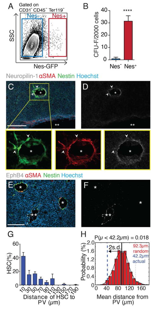
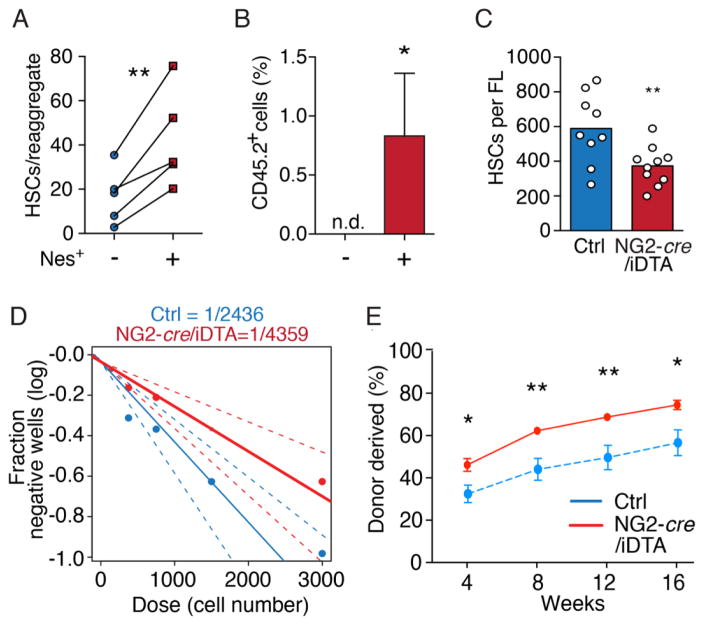
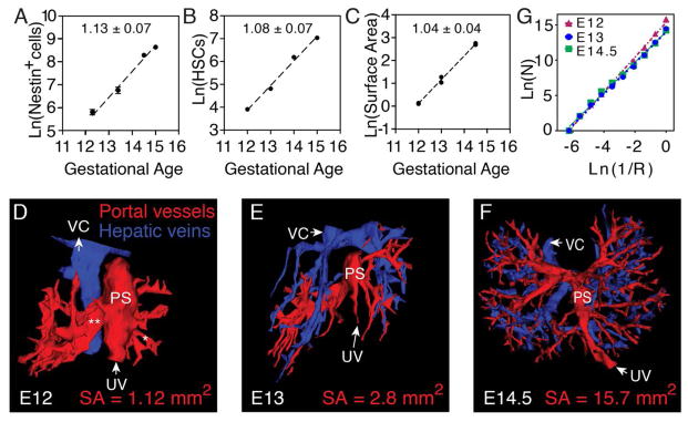
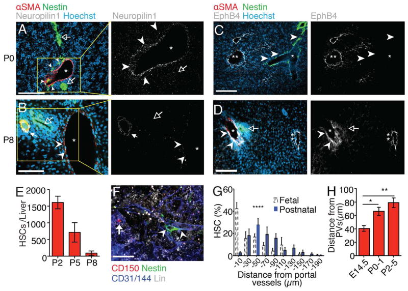
Whereas the cellular basis of the hematopoietic stem cell (HSC) niche in the bone marrow has been characterized, the nature of the fetal liver niche is not yet elucidated. We show that Nestin(+)NG2(+) pericytes associate with portal vessels, forming a niche promoting HSC expansion. Nestin(+)NG2(+) cells and HSCs scale during development with the fractal branching patterns of portal vessels, tributaries of the umbilical vein. After closure of the umbilical inlet at birth, portal vessels undergo a transition from Neuropilin-1(+)Ephrin-B2(+) artery to EphB4(+) vein phenotype, associated with a loss of periportal Nestin(+)NG2(+) cells and emigration of HSCs away from portal vessels. These data support a model in which HSCs are titrated against a periportal vascular niche with a fractal-like organization enabled by placental circulation.


Conflict of interest statement

The authors declare no competing financial interests.

Figures

Fig. 1
Fig. 1. Peri-arterial Nestin+ stromal cells associate with HSCs in the fetal liver
(A) FACS analysis of Nestin-GFP stroma from E14.5 FL. (B) CFU-F from sorted FL cells, n = 4. (C to F) Immunofluorescence analyses of Nestin-GFP E14.5 FL cryosections stained for Neuropilin-1 (white; C, D) or EphB4 (white; E, F). Colocalization of αSMA+ and Nestin+ pericytes around Neuropilin-1+ portal vessels (*) but not EphB4+ veins (**). Scale bar, 100 μm. (G) Distance distribution between CD150+CD48CD41Lineage HSCs and Nestin+ cells from whole-mount stained Nestin-GFP FL (n = 105 from 12 E14.5 FLs), binned into 20 μm intervals. (H) Probability distribution of mean distances from simulations of randomly positioned HSCs in relation to Nestin+ cells. Actual mean distance shown in relation to mean of simulations ± 2 s.d. ****P < 10−4.
Fig. 2
Fig. 2. Nestin+ perivascular cells drive HSC expansion in vivo
(A) FACS quantification of CD150+Sca1+CD45+CD48Lin HSCs in reaggregates with or without Nestin+ cells. (B) Donor engraftment 16 weeks after transplantation of reaggregates together with congenic competitor cells. n.d. = not detectable. n = 6–7 per group. (C) CD150+CD48Sca1+CD11b+CD45+CD41LineageDAPI HSC numbers in control and NG2-depleted littermates. (D) Limiting dilution analyses of LTC-ICs in Lineage cells from NG2-depleted (red) and control (blue) littermate FLs. (E) Competitive repopulation assays of NG2-depleted and control FL. n = 6/7 mice per control/depleted littermates. *P < 0.05, **P < 0.01.
Fig. 3
Fig. 3. Fractal geometry underlies HSC expansion
(A) Nestin+ cells expand from E12–E15 according to a power law (R2 = 0.95). (B) HSC numbers relative to gestational age plotted from quantification of Ema et al. (5). (C) Portal vessel surface area from E12–E14.5 scales according to similar power law (R2 = 0.99; n = 6). Slopes indicated in panels. (D to F) 3D reconstructions of afferent PVs (red) and efferent veins (blue) in E12 (D), E13 (E), and E14.5 (F) FLs. PS, portal sinus; VC, vena cava; SA, surface area. (G) Calculation of the scale-invariant fractal dimension of FL afferent vessels using box-counting.
Fig. 4
Fig. 4. Ligation of the umbilical vein inlet terminates the portal vessel HSC niche
(A to D) Immunofluorescence analyses of postnatal day 0 (P0; A and C) and P8 (B and D) liver cryosections stained for αSMA (red), Nestin-GFP (green), nuclear dye Hoechst (blue), and Neuropilin-1 or EphB4 (white). *portal vessels; **hepatic veins. Arrowheads: Nestin+ cells (P0) or outline of portal vessel; Solid arrow: hepatic artery, open arrows: bile ducts. (E) FACS analysis showing decreasing numbers of CD150+CD48Sca1+CD11b+CD45+CD41LineageDAPI HSCs between P2 and P8. (F) Representative whole-mount immunofluorescence imaging of a CD150+CD48Lin HSC (arrow) in P3 fetal liver. Arrowheads: portal vessel. (G) Distance distribution of HSCs from portal vessels (n = 77 from 20 livers). FL HSC measurements from Fig. 1G (dashed-line bars) are shown for comparison. (H) HSC distances from portal vessels in fetal and postnatal livers showing increasing mean distances with age. Scale bars: A–D, 100 μm, F, 20 μm. *P < 0.05, **P < 0.01, ****P < 0.0001.

Comment in

  • STEM CELLS. Potency finds its niches.
    Cabezas-Wallscheid N, Trumpp A. Cabezas-Wallscheid N, et al. Science. 2016 Jan 8;351(6269):126-7. doi: 10.1126/science.aae0325. Science. 2016. PMID: 26744396 No abstract available.

References

    1. Medvinsky A, Dzierzak E. Definitive hematopoiesis is autonomously initiated by the AGM region. Cell. 1996;86:897–906. doi: 10.1016/S0092-8674(00)80165-8. - DOI - PubMed
    1. Boisset JC, van Cappellen W, Andrieu-Soler C, Galjart N, Dzierzak E, Robin C. In vivo imaging of haematopoietic cells emerging from the mouse aortic endothelium. Nature. 2010;464:116–120. doi: 10.1038/nature08764. - DOI - PubMed
    1. de Bruijn MF, Speck NA, Peeters MC, Dzierzak E. Definitive hematopoietic stem cells first develop within the major arterial regions of the mouse embryo. EMBO J. 2000;19:2465–2474. doi: 10.1093/emboj/19.11.2465. - DOI - PMC - PubMed
    1. Gekas C, Dieterlen-Lièvre F, Orkin SH, Mikkola HKA. The placenta is a niche for hematopoietic stem cells. Dev Cell. 2005;8:365–375. doi: 10.1016/j.devcel.2004.12.016. - DOI - PubMed
    1. Ema H, Nakauchi H. Expansion of hematopoietic stem cells in the developing liver of a mouse embryo. Blood. 2000;95:2284–2288. - PubMed

Publication types