. 2015 Dec 18;350(6267):1548-51.
doi: 10.1126/science.aad8305. Epub 2015 Dec 3.

Structure and membrane remodeling activity of ESCRT-III helical polymers

Affiliations

Structure and membrane remodeling activity of ESCRT-III helical polymers

John McCullough et al. Science. .

Abstract

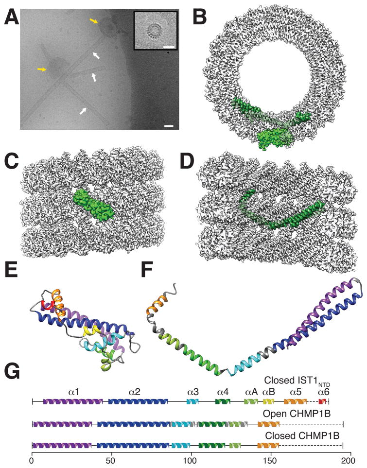
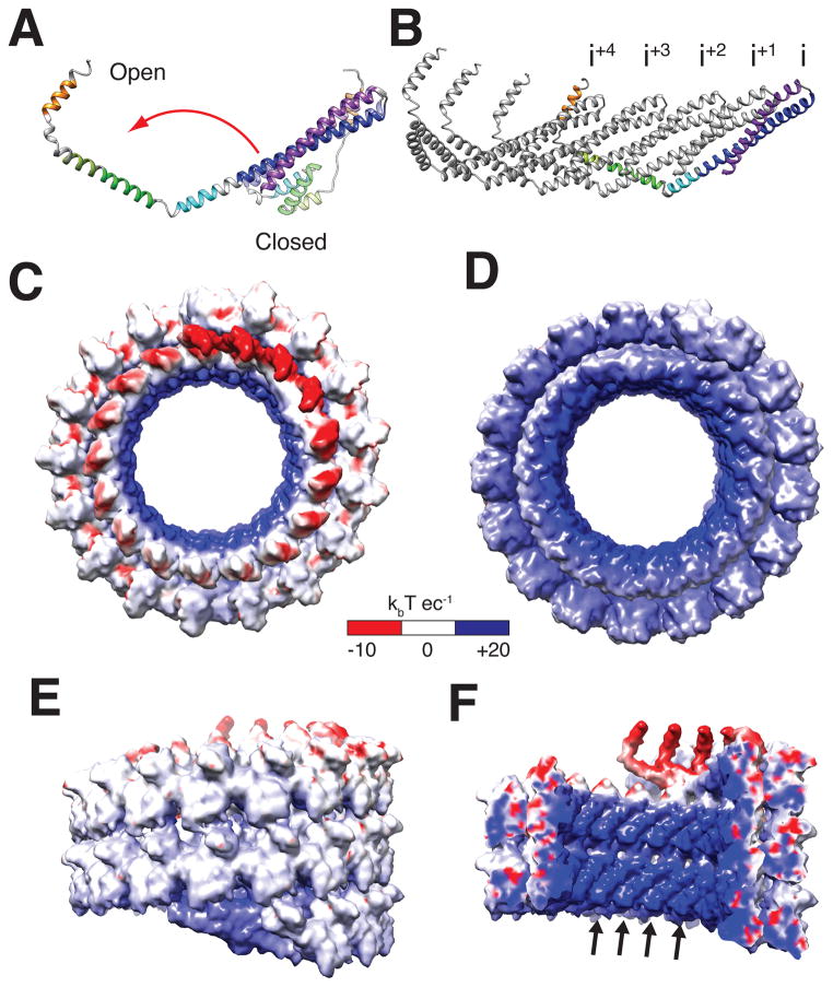
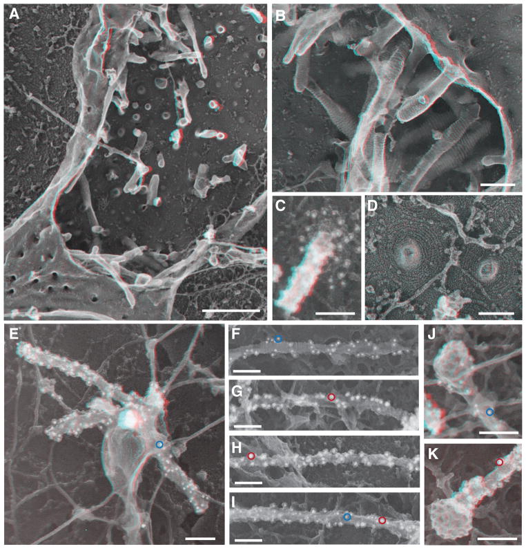
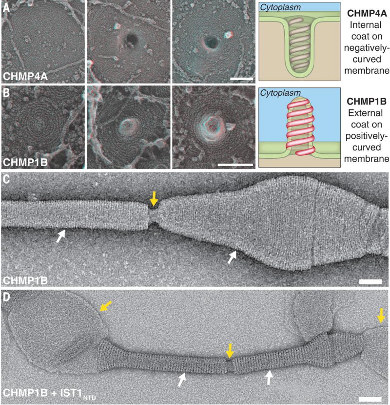
The endosomal sorting complexes required for transport (ESCRT) proteins mediate fundamental membrane remodeling events that require stabilizing negative membrane curvature. These include endosomal intralumenal vesicle formation, HIV budding, nuclear envelope closure, and cytokinetic abscission. ESCRT-III subunits perform key roles in these processes by changing conformation and polymerizing into membrane-remodeling filaments. Here, we report the 4 angstrom resolution cryogenic electron microscopy reconstruction of a one-start, double-stranded helical copolymer composed of two different human ESCRT-III subunits, charged multivesicular body protein 1B (CHMP1B) and increased sodium tolerance 1 (IST1). The inner strand comprises "open" CHMP1B subunits that interlock in an elaborate domain-swapped architecture and is encircled by an outer strand of "closed" IST1 subunits. Unlike other ESCRT-III proteins, CHMP1B and IST1 polymers form external coats on positively curved membranes in vitro and in vivo. Our analysis suggests how common ESCRT-III filament architectures could stabilize different degrees and directions of membrane curvature.


Figures

Fig. 1
Fig. 1. IST1NTD and CHMP1B copolymerized into helical tubes comprising polar, double-stranded helical filaments
(A) Electron cryomicrograph showing IST1NTD-CHMP1B tubes (white arrows) assembled by incubating equimolar IST1NTD and CHMP1B in the presence of polymer-nucleating small acidic unilamellar vesicles (SUVs, yellow arrows). Inset: end-on view of a short IST1NTD-CHMP1B tube. Bars: 40 nm (A), 20 nm (inset). (B) End-on view of the reconstructed IST1NTD-CHMP1B tube highlighting single subunits of IST1NTD (light green, outer strand) and CHMP1B (dark green, inner strand). (C) External view of the reconstructed helix with a highlighted IST1NTD subunit. (D) Internal cutaway view of the reconstructed helix with a highlighted CHMP1B subunit. (E) Ribbon diagram of the modeled IST1NTD subunit (closed conformation). (F) Ribbon diagram of the modeled CHMP1B subunit (open conformation). (G) Secondary structure diagrams for closed IST1NTD (top), open CHMP1B (middle), and closed CHMP1B (bottom).
Fig. 2
Fig. 2. CHMP1B opening, strand structure, and electrostatic surface potentials of the IST1NTD-CHMP1B assembly
(A) Superposition of the open and closed CHMP1B conformations. (B) Five interlocked CHMP1B molecules from the inner strand of the filament. (C) “Top-end”, electrostatic surface view of the IST1NTD-CHMP1B tube, highlighting the acidity of the CHMP1B inner strand (including Glu130, Asp131, Asp147, Glu152, Asp155, Glu156, Asp160) and the IST1NTD outer strand (including Asp49, Glu50, Glu57, Glu163, Glu168, Glu178, Asp180, Glu186). (D) “Bottom-end”, electrostatic surface view of the IST1NTD-CHMP1B tube, highlighting the strongly basic characters of the CHMP1B inner strand (including Lys3, Lys87, Lys94, Lys101, Lys107, and Lys114) and the IST1NTD outer strand (including Lys7, Arg10, Lys90, Arg109, Lys118, Lys127, Lys130, Lys134, Arg137). (E) Exterior, electrostatic surface view of the IST1NTD-CHMP1B tube, revealing the modestly basic character of the IST1NTD outer strand. (F) Internal cutaway electrostatic surface view of the IST1NTD-CHMP1B tube, revealing the strongly basic character of the lumenal surface, contributed primarily by basic residues in CHMP1B helix 1 (arrows), including Lys3, Lys13, Arg17, Lys20, Lys21, Lys24, Lys32, and Lys35.
Fig. 3
Fig. 3. CHMP1B and IST1 tubulated cellular membranes
(A) Survey view of the cytoplasmic surface of the plasma membrane in an unroofed COS-7 cell expressing FLAG-CHMP1B. Tubular invaginations extending into the cell interior are apparent along the exposed plasma membrane and as stabilized openings at the edges of the cell. Use view glasses for 3D viewing of anaglyphs (left eye = red). (B) Higher magnification view of tubular invaginations induced and coated by FLAG-CHMP1B filaments. (C) Immunodecoration confirmed the presence of CHMP1B around and along a tubule in a cell expressing untagged CHMP1B. Antibody detected with 12 nm gold is white in these contrast reversed EM images. (D) Higher magnification view of FLAG-CHMP1B filament spirals on exposed plasma membrane. (E) CHMP1B-immunoreactive organelle in an unroofed COS-7 cell expressing untagged CHMP1B. Antibody detected with 12 nm gold is white in these contrast reversed EM images; a representative gold particle is circled in blue. (F to I) Representative internal tubules from cells co-expressing untagged CHMP1B (12 nm gold, example circled in blue) and IST1-myc (18 nm gold); examples circled in red. (J and K) Clathrin coated bud capping the end of CHMP1B (J) and IST1-myc (K) immuno-labeled tubules from co-transfected cells. Importantly, measurements of filament diameter (and interstrand spacing) showed that when apparently unitary filaments were resolvable, their diameter varied from 5–10 nm including platinum. These measurements are generally consistent with the dimensions of IST1-CHMP1B and CHMP1B filaments formed in vitro. Scale bars 500nm (A), 100nm (B) to (K).
Fig. 4
Fig. 4. Topology of ESCRT-III membrane deformation in cells and in vitro
(A) Series of filament spirals on the plasma membrane of COS-7 cells expressing CHMP4A1–164 show development of the outwardly directed protrusions previously associated with ESCRT-III filaments (15, 16). Drawing highlights relationship between CHMP4A filament spiral and a negatively-curved plasma membrane tubule. (B) Series of filament spirals on the plasma membrane of COS-7 cells expressing FLAG-CHMP1B show development of invaginations directed into the cell. Drawing highlights relationship between CHMP1B filament spiral and a positively-curved plasma membrane tubule. (C) Negative stain electron micrograph showing that CHMP1B tubulates liposomes and forms a filamentous coat on the outside of the tubule. White arrows highlight regions coated by the CHMP1B helices, and the yellow arrow highlights a break in the coat where the internal lipid is visible. (D) Negative stain electron micrograph showing that the IST1NTD-CHMP1B copolymer forms on the outside of membrane tubules. White arrows highlight regions coated by the IST1NTD-CHMP1B copolymer, and the yellow arrows highlight breaks in the helical coat or uncoated regions of the liposome where the internal membrane is visible. Scale bars 100nm (A) and (B), 50 nm (C) and (D).

References

    1. Hurley JH. ESCRTs are everywhere. EMBO J. 2015;34:2398–2407. doi: 10.15252/embj.201592484. - DOI - PMC - PubMed
    1. Hanson PI, Cashikar A. Multivesicular body morphogenesis. Annu Rev Cell Dev Biol. 2012;28:337–362. doi: 10.1146/annurev-cellbio-092910-154152. - DOI - PubMed
    1. McCullough J, Colf LA, Sundquist WI. Membrane fission reactions of the mammalian ESCRT pathway. Annu Rev Biochem. 2013;82:663–692. doi: 10.1146/annurev-biochem-072909-101058. - DOI - PMC - PubMed
    1. Bajorek M, Schubert HL, McCullough J, Langelier C, Eckert DM, Stubblefield WM, Uter NT, Myszka DG, Hill CP, Sundquist WI. Structural basis for ESCRT-III protein autoinhibition. Nat Struct Mol Biol. 2009;16:754–762. doi: 10.1038/nsmb.1621. - DOI - PMC - PubMed
    1. Muzioł T, Pineda-Molina E, Ravelli RB, Zamborlini A, Usami Y, Göttlinger H, Weissenhorn W. Structural basis for budding by the ESCRT-III factor CHMP3. Dev Cell. 2006;10:821–830. doi: 10.1016/j.devcel.2006.03.013. - DOI - PubMed

Publication types

MeSH terms

Associated data