. 2016 Jan;26(1):119-30.
doi: 10.1038/cr.2015.143. Epub 2015 Dec 4.

Genetic lineage tracing identifies in situ Kit-expressing cardiomyocytes

Affiliations

Genetic lineage tracing identifies in situ Kit-expressing cardiomyocytes

Qiaozhen Liu et al. Cell Res. 2016 Jan.

Abstract

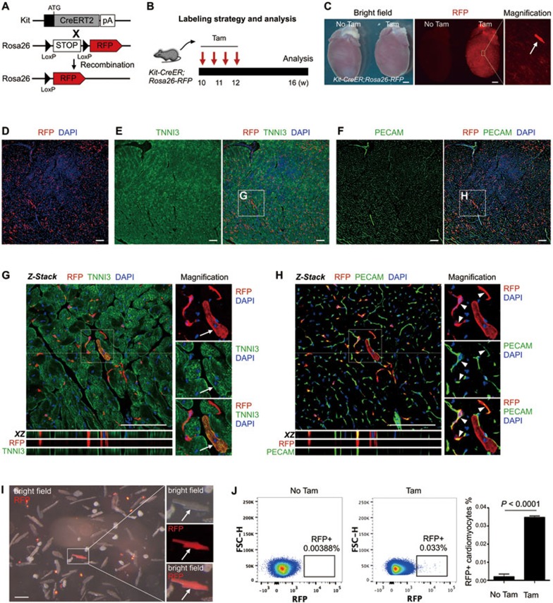
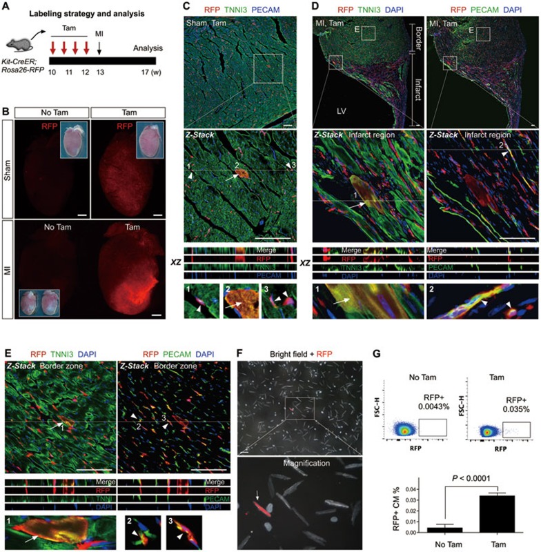
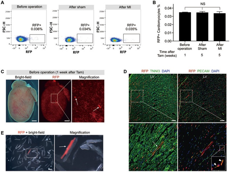
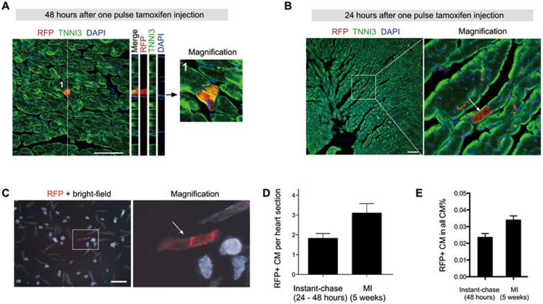
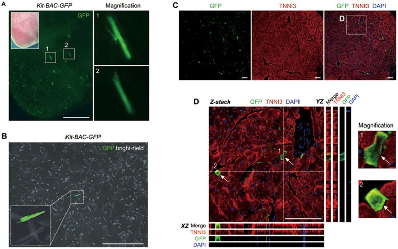
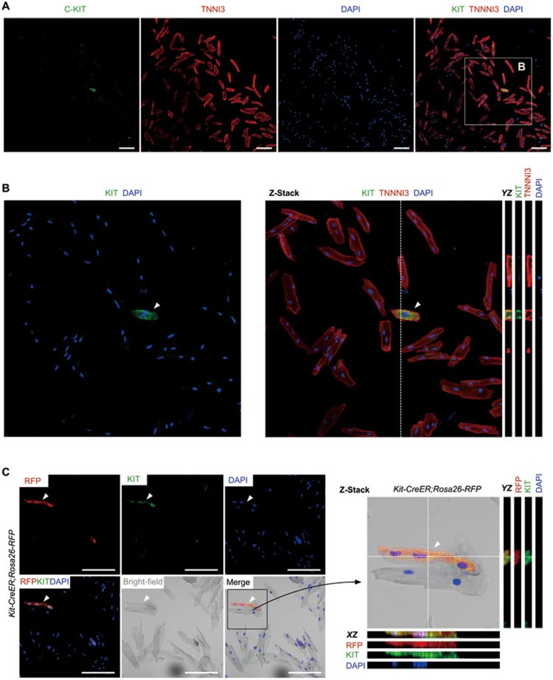
Cardiac cells marked by c-Kit or Kit, dubbed cardiac stem cells (CSCs), are in clinical trials to investigate their ability to stimulate cardiac regeneration and repair. These studies were initially motivated by the purported cardiogenic activity of these cells. Recent lineage tracing studies using Kit promoter to drive expression of the inducible Cre recombinase showed that these CSCs had highly limited cardiogenic activity, inadequate to support efficient cardiac repair. Here we reassess the lineage tracing data by investigating the identity of cells immediately after Cre labeling. Our instant lineage tracing approach identifies Kit-expressing cardiomyocytes, which are labeled immediately after tamoxifen induction. In combination with long-term lineage tracing experiments, these data reveal that the large majority of long-term labeled cardiomyocytes are pre-existing Kit-expressing cardiomyocytes rather than cardiomyocytes formed de novo from CSCs. This study presents a new interpretation for the contribution of Kit(+) cells to cardiomyocytes and shows that Kit genetic lineage tracing over-estimates the cardiogenic activity of Kit(+) CSCs.


Figures

Figure 1
Figure 1
The fate of Kit+ cells in heart homeostasis. (A) Kit-CreER mice were crossed with Rosa26-RFP mice for lineage tracing. (B) A schematic of tamoxifen induction and analysis. (C) Whole mount bright field and fluorescence view of Kit-CreER;Rosa26-RFP hearts with or without tamoxifen (Tam or No Tam). Arrow indicates RFP+ cardiomyocyte. (D-F) Immunostaining for RFP, TNNI3 and PECAM on Kit-CreER;Rosa26-RFP heart sections. (G, H) Z-stack confocal images including XZ scanned sections showing RFP+TNNI3+ cells (arrows, G) and RFP+PECAM+ cells (arrowheads, H). (I) Image of cells dissociated from hearts of Kit-CreER;Rosa26-RFP mice. Arrow indicates a rare RFP+ cardiomyocyte. (J) Quantification of RFP+ cardiomyocytes by flow cytometry. Events in images are gated on live and lineage-negative cardiomyocytes. n = 4. Scale bars represent 1 mm in C, 100 μm in D-I.
Figure 2
Figure 2
The fate of Kit+ cells after MI. (A) A schematic of tamoxifen induction, MI and analysis. (B) Whole mount fluorescence view of sham or MI hearts with or without tamoxifen treatment (Tam or No Tam). Inserts are the same hearts in bright field. (C) Immunostaining for RFP, TNNI3 and PECAM on Kit-CreER;Rosa26-RFP heart sections (Sham, Tam). Kit-CreER labels cardiomyocytes (RFP+TNNI3+, arrow) or endothelial cells (RFP+PECAM+, arrowheads). (D, E) Immunostaining for RFP, TNNI3 and PECAM on heart sections after MI. Kit-CreER labels cardiomyocytes (RFP+TNNI3+, arrow) or endothelial cells (RFP+PECAM+, arrowheads) in both infarcted (D) and border regions (E). Z-stack images were taken for all magnified regions. XZ scanned images are shown by merge and split channels. 1, 2, 3 are magnified images of cells indicated by arrows or arrowheads. (F) Images of cells dissociated from MI hearts of Kit-CreER;Rosa26-RFP mice. Arrow indicates a rare RFP+ cardiomyocyte. (G) Quantification of the percentage of RFP+ cardiomyocytes in MI hearts by flow cytometry. n = 4. Scale bars, 1 mm in B; 100 μm in C-F.
Figure 3
Figure 3
Quantification of Kit-CreER-labeled cardiomyocytes before and after MI. (A) Flow cytometric analysis of RFP+ cardiomyocytes isolated from Kit-CreER;Rosa26-RFP mouse hearts. (B) Quantification of the percentage of RFP+ cardiomyocytes labeled by Kit-CreER. n = 4; NS, non-significant. (C) Whole mount bright field and fluorescence view of Kit-CreER;Rosa26-RFP mouse heart. Arrows indicate RFP+ cardiomyocytes. (D) Immunostaining for RFP, TNNI3 and PECAM in heart sections reveals RFP+ cardiomyocyte (arrow) and endothelial cells (arrowheads). LV, left ventricle. (E) Image of cells dissociated from Kit-CreER;Rosa26-RFP mouse hearts (n = 3 hearts analyzed). Arrow indicates a RFP+ cardiomyocyte. Scale bars, 1 mm in C, 100 μm in D and E.
Figure 4
Figure 4
Instant lineage tracing reveals Kit-expressing cardiomyocytes. (A) Z-stack confocal images showing RFP and TNNI3 in heart sections from Kit-CreER;Rosa26-RFP mice at 48 h after tamoxifen injection. (B) Immunostaining for RFP and TNNI3 in heart sections from mice collected 24 h after tamoxifen injection. (C) Images of heart cells isolated from mice at 24 h after tamoxifen injection. Arrow indicates the RFP+ cardiomyocyte. (D) Quantification of RFP+ cardiomyocytes (CM) per heart sections in instant lineage tracing heart samples (24-48 h after tamoxifen treatment) versus hearts after MI (5 weeks after tamoxifen treatment). For each heart, 80 to 100 slides were examined for quantification, and 4-6 hearts from each group were analyzed. (E) Quantification of the percentage of RFP+ cardiomyocytes by flow cytometry. n = 4. Scale bar, 100 μm.
Figure 5
Figure 5
Kit is in situ expressed by cardiomyocytes in the adult heart. (A) Whole mount fluorescence view of the adult Kit-BAC-GFP heart. Boxed regions are magnified in right panels. GFP is a surrogate for Kit. (B) Image of isolated cells from the adult Kit-BAC-GFP heart shows Kit+ cardiomyocytes. (C) Immunostaining for GFP and TNNI3 on heart sections of Kit-BAC-GFP mice. (D) Z-stack confocal images along the XZ and YZ axes. 1 and 2 denote two Kit+ cardiomyocytes (arrows). Images are representatives of three individual samples. Scale bars, 1 mm in A and B, 100 μm in C and D.
Figure 6
Figure 6
Kit is expressed by cardiomyocytes. (A) Immunostaining for Kit and TNNI3 on cardiomyocytes isolated from wild type heart. (B) Averaged intensity from projection of 9 optical sections; YZ sections (from dotted lines) are also shown. Arrowhead indicates Kit+ cardiomyocyte. (C) Immunostaining for RFP and Kit on cardiomyocytes isolated from Kit-CreER;Rosa26-RFP mice treated with tamoxifen. Averaged intensity from projection of 9 optical sections; XZ and YZ sections (from dotted lines) are also shown. Scale bar, 100 μm.

References

    1. Anversa P, Kajstura J, Rota M, Leri A. Regenerating new heart with stem cells. J Clin Invest 2013; 123:62–70. - PMC - PubMed
    1. Garbern JC, Lee RT. Cardiac stem cell therapy and the promise of heart regeneration. Cell Stem Cell 2013; 12:689–698. - PMC - PubMed
    1. Loffredo FS, Steinhauser ML, Gannon J, Lee RT. Bone marrow-derived cell therapy stimulates endogenous cardiomyocyte progenitors and promotes cardiac repair. Cell Stem Cell 2011; 8:389–398. - PMC - PubMed
    1. Chugh AR, Beache GM, Loughran JH, et al. Administration of cardiac stem cells in patients with ischemic cardiomyopathy: the SCIPIO trial: surgical aspects and interim analysis of myocardial function and viability by magnetic resonance. Circulation 2012; 126:S54–S64. - PMC - PubMed
    1. Bolli R, Chugh AR, D'Amario D, et al. Cardiac stem cells in patients with ischaemic cardiomyopathy (SCIPIO): initial results of a randomised phase 1 trial. Lancet 2011; 378:1847–1857. - PMC - PubMed

Publication types

Substances